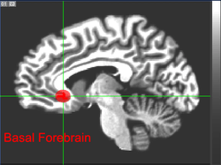
Basal forebrain
| Basal forebrain | |
|---|---|
 The Basal Forebrain | |
| Details | |
| Identifiers | |
| Latin | pars basalis telencephali |
| NeuroNames | ancil-1997 |
| NeuroLex ID | Basal Forebrain |
| TA | A14.1.09.401 |
| FMA | 77700 |
The basal forebrain is a collection of structures located to the front of and below the striatum. It includes the nucleus accumbens, nucleus basalis, diagonal band of Broca, substantia innominata, and medial septal nuclei. These structures are important in the production of acetylcholine, which is then distributed widely throughout the brain. The basal forebrain is considered to be the major cholinergic output of the central nervous system (CNS).
Function
Acetylcholine is known to promote wakefulness in the basal forebrain. Stimulating the basal forebrain gives rise to acetylcoline release, which induces wakefulness and REM sleep, whereas inhibition of acetylcholine release in the basal forebrain by adenosine causes slow wave sleep.
Adenosine acts on A1 receptors of cholinergic neurons in the basal forebrain. This results in hyperpolarization of cholinergic neurons, which inhibits the release of acetylcholine.
The basal forebrain and adjacent areas are a focus for sleep research. Research, conducted by investigators from Children's Hospital Boston and the University of Helsinki, ties together previous observations about sleep and finds that nitric oxide production in the basal forebrain is both necessary and sufficient to produce sleep.[1]
Clinical significance
Acetylcholine affects the ability of brain cells to transmit information to one another, and also encourages neuronal plasticity, or learning. Thus, damage to the basal forebrain can reduce the amount of acetylcholine in the brain and impair learning. This may be one reason why basal forebrain damage can result in memory impairments such as amnesia and confabulation. One common cause of basal forebrain damage is aneurysm of the anterior communicating artery.[2]