Chylothorax
| Chylothorax | |
|---|---|
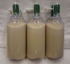 | |
| Milky fluid removed from the chest of a patient with a chylothorax | |
| Classification and external resources | |
| Specialty | pulmonology |
| ICD-10 | I89.8, J91 |
| ICD-9-CM | 457.8 |
| DiseasesDB | 29612 |
| eMedicine | med/381 |
| MeSH | D002916 |
A chylothorax (or chyle leak) is a type of pleural effusion. It results from lymph formed in the digestive system called chyle accumulating in the pleural cavity due to either disruption or obstruction of the thoracic duct.
In people on a normal diet, this effusion can be identified by its turbid, milky white appearance, since chyle contains high levels of triglycerides. It is important to distinguish chylothorax from pseudochylothorax (pleural effusions high in cholesterol), which has a similar appearance, but is caused by more chronic inflammatory processes, and has a different treatment. [1]
Causes
The condition is rare but serious, and appears in all mammals. It results from leakage of lymph fluid from the thoracic duct (or one of its tributaries). This can result from direct laceration (e.g., from surgery) or from nontraumatic causes. The most common nontraumatic cause is malignancy, especially lymphoma. Less common is left-heart failure, infections, and developmental abnormalities such as Down syndrome and Noonan syndrome.[2]
Treatment
Since the mechanism behind chylothorax is not well understood, treatment options are limited. Drainage of the fluid out of the pleural space is essential to obviate damage to organs, especially the inhibition of lung function by the counter pressure of the chyle. Another treatment option is pleuroperitoneal shunting (creating a communication channel between pleural space and peritoneal cavity). By this surgical technique loss of essential triglycerides that escape the thoracic duct can be prevented. Omitting fat (in particular FFA) from the diet is essential. Either surgical or chemical pleurodesis are options: the leaking of lymphatic fluids is stopped by irritating the lungs and chest wall, resulting in a sterile inflammation. This causes the lung and the chest wall to be fused together which prevents the leaking of lymphatic fluids into the pleural space. The medication octreotide has been shown to be beneficial and in some cases will stop the chylothorax after a few weeks.[3][4]
In animals, the most effective form of treatment until recently has been surgical ligation of the thoracic duct combined with partial pericardectomy.[5] There is at least one case report (in a cat) of clinical response to treatment with rutin.[6]
See also
References
- ↑ Heffner, John. "Clinical presentation, diagnosis and management of cholesterol effusions". 8.0. UpToDate. Retrieved 30 October 2014.
- ↑
- ↑ Dalokay Kilic, MD; Ekber Sahin, MD; Oner Gulcan, MD; Bulent Bolat, MD; Riza Turkoz, MD; Ahmet Hatipoglu, MD (2005). "Octreotide for Treating Chylothorax after Cardiac Surgery". Texas Heart Institute Journal. 32 (3): 437–39. PMC 1336729
 . PMID 16392238.
 - ↑ Marcia L. Buck, Pharm.D., FCCP (2004). "Octreotide for the Management of Chylothorax in Infants and Children". Pediatric Pharmacotherapy. 10 (10).
- ↑ Birchard SJ, Smeak DD, McLoughlin MA (March 1998). "Treatment of idiopathic chylothorax in dogs and cats". J. Am. Vet. Med. Assoc. 212 (5): 652–7. PMID 9524635.
- ↑ Kopko SH (August 2005). "The use of rutin in a cat with idiopathic chylothorax". Can. Vet. J. 46: 729–31. PMC 1180424. PMID 16187718.
External links
- University of Wisconsin: http://www.vetmed.wisc.edu/dss/ChyloThoraxTrial/page5.php
- Morris Animal Foundation: http://www.morrisanimalfoundation.org/ada_studies.php?col=study&val=179