Colles' fracture
| Colles' fracture | |
|---|---|
| Classification and external resources | |
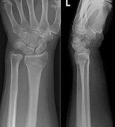
 An X-ray image of a fractured radius showing the characteristic Colles' fracture with displacement and angulation of the distal end of the radius | |
| ICD-10 | S52.5 |
| AO | 23-A2.2 |
| MeSH | 68003100 |
A Colles' fracture is a fracture of the distal radius in the forearm with dorsal (posterior) and radial displacement of the wrist and hand.[1] The fracture is sometimes referred to as a "dinner fork" or "bayonet" deformity due to the shape of the resultant forearm. Colles' fractures are often seen in people with osteoporosis.
Terminology
The term of Colles fracture is classically used to describe a fracture at the distal end of the radius, at its cortico-cancellous junction. However, now the term tends to be used loosely to describe any fracture of the distal radius, with or without involvement of the ulna, that has dorsal displacement of the fracture fragments. Colles himself described it as a fracture that “takes place at about an inch and a half (38mm) above the carpal extremity of the radius” and “the carpus and the base of metacarpus appears to be thrown backward”.[2]
The classic Colles fracture has the following characteristics:[3]
- Transverse fracture of the radius
- 2.5 cm (0.98 inches) proximal to the radio-carpal joint
- dorsal displacement and dorsal angulation, together with radial tilt[1]
Causes
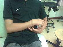
The fracture is most commonly caused by people falling onto a hard surface and breaking their fall with outstretched arms - falling with wrists flexed would lead to a Smith's fracture. It can also be caused by overuse. Originally it was described in elderly and/or post-menopausal women. It usually occurs about three to five centimetres proximal to the radio-carpal joint with posterior and lateral displacement of the distal fragment resulting in the characteristic "dinner fork" or "bayonet" like deformity. Colles' fracture is a common fracture in people with osteoporosis, second only to vertebral fractures.
Diagnosis


- Dorsal tilt
- Radial shortening
- Loss of ulnar inclination≤
- Radial angulation of the wrist
- Dorsal displacement of the distal fragment
- Comminution at the fracture site
- Associated fracture of the ulnar styloid process in more than 60% of cases.
Treatment
Management depends on the severity of the fracture. An undisplaced fracture may be treated with a cast alone. The cast is applied with the distal fragment in palmar flexion and ulnar deviation. A fracture with mild angulation and displacement may require closed reduction. There is some evidence that immobilization with the wrist in dorsiflexion as opposed to palmarflexion results in less redisplacement and better functional status.[4] Significant angulation and deformity may require an open reduction and internal fixation or external fixation. The volar forearm splint is best for temporary immobilization of forearm, wrist and hand fractures, including Colles' fracture. There are several established instability criteria: dorsal tilt >20°, comminuted fracture, abruption of the ulnar styloid process, intraarticular displacement >1mm, loss of radial height >2mm.
A higher amount of instability criteria increases the likelihood of operative treatment.
Treatment modalities differ in the elderly.[5]
Eponym
It is named after Abraham Colles (1773–1843), an Irish surgeon, from Kilkenny. who first described it in 1814 by simply looking at the classical deformity before the advent of X-rays.[6] Ernest Amory Codman was the first to study it using X-rays. His article, published in the Boston Medical and Surgical Journal, now known as The New England Journal of Medicine, also developed the classification system.[7][8]
In older and younger people
Colles fractures occur in all age groups, although certain patterns follow an age distribution.
- In the elderly, because of the weaker cortex, the fracture is more often extra-articular.
- Younger individuals tend to require a higher energy force to cause the fracture and tend to have more complex intra-articular fractures. In children with open epiphyses, an equivalent fracture is the "epiphyseal slip", as can be seen in other joints, such as a slipped capital femoral epiphysis in the hip. This is a Salter I or II fracture with the deforming forces directed through the weaker epiphyseal plate.
- More common in women because of post-menopausal osteoporosis.
See also
References
- 1 2 Solomon et al., Apley's system of orthopaedics and fractures, 9th ed., p.772
- ↑ Colles A 2006 On the fracture of the carpal extremity of the radius. Edinb Med Surg J. 1814;10:181. Clin Orthop Relat Res 445:5-7.
- ↑ GP Notebook. "Colles' fracture". Retrieved 2009-02-21.
- ↑ "Adult Distal Radius Frx: Non Operative Treatment - Wheeless' Textbook of Orthopaedics".
- ↑ Blakeney, William (November 2010). "Stabilization and treatment of Colles’ fractures in elderly patients". Clinical Interventions in Aging: 337. doi:10.2147/CIA.S10042.
- ↑ synd/2152 at Who Named It?
- ↑ Mallon, Bill (2000). Ernest Amory Codman : the end result of a life in medicine. Philadelphia: Saunders. ISBN 978-0-7216-8461-1.
- ↑ CODMAN, E. A. (1900). "A Study of the X-Ray Plates of One Hundred and Forty Cases of Fracture of the Lower End of the Radius". The Boston Medical and Surgical Journal. 143 (13): 305–308. doi:10.1056/NEJM190009271431301. ISSN 0096-6762.
External links
- Colles Fracture Detailed information for Orthopedic Doctors