Denaturation (biochemistry)

Process of partial or total alteration of the native secondary, and/or tertiary, and/or quaternary structures of proteins or nucleic acids resulting in a loss of bioactivity.
Note 1: Modified from the definition given in ref.[1]
Note 2: Denaturation can occur when proteins and nucleic acids are subjected to elevated temperature or to extremes of pH, or to nonphysiological concentrations of salt, organic solvents, urea, or other chemical agents.
Note 3: An enzyme loses its catalytic activity when it is denaturized.[2]
Denaturation is a process in which proteins or nucleic acids lose the quaternary structure, tertiary structure and secondary structure which is present in their native state, by application of some external stress or compound such as a strong acid or base, a concentrated inorganic salt, an organic solvent (e.g., alcohol or chloroform), radiation or heat.[3] If proteins in a living cell are denatured, this results in disruption of cell activity and possibly cell death. Protein denaturation is also a consequence of cell death.[4][5] Denatured proteins can exhibit a wide range of characteristics, from conformational change and loss of solubility to aggregation due to the exposure of hydrophobic groups.
Protein folding is key to whether a globular protein or a membrane protein can do its job correctly. It must be folded into the right shape to function. But hydrogen bonds, which play a big part in folding, are rather weak, and it doesn't take much heat, acidity, varying salt concentrations, or other stress to break some and form others, denaturing the protein. This is one reason why tight homeostasis is physiologically necessary in many life forms.
This concept is unrelated to denatured alcohol, which is alcohol that has been mixed with additives to make it unsuitable for human consumption.
Common examples

When food is cooked, some of its proteins become denatured. This is why boiled eggs become hard and cooked meat becomes firm.
A classic example of denaturing in proteins comes from egg whites, which are typically largely egg albumins in water. Fresh from the eggs, egg whites are transparent and liquid. Cooking the thermally unstable whites turns them opaque, forming an interconnected solid mass. The same transformation can be effected with a denaturing chemical. Pouring egg whites into a beaker of acetone will also turn egg whites translucent and solid. The skin that forms on curdled milk is another common example of denatured protein. The cold appetizer known as ceviche is prepared by chemically "cooking" raw fish and shellfish in an acidic citrus marinade, without heat.[6]
Protein denaturation
Denatured proteins can exhibit a wide range of characteristics, from loss of solubility to protein aggregation."
1) Primary Structure : the linear structure of amino acids in the polypeptide chain
2) Secondary Structure : hydrogen bonds between peptide group chains in an alpha helix or beta sheet
3) Tertiary Structure : three-dimensional structure of alpha helixes and beta helixes folded
4) Quaternary Structure : three-dimensional structure of multiple polypeptides and how they fit together

Background
Proteins are amino acid polymers. A protein is created by ribosomes that "read" RNA that is encoded by codons in the gene and assemble the requisite amino acid combination from the genetic instruction, in a process known as translation. The newly created protein strand then undergoes posttranslational modification, in which additional atoms or molecules are added, for example copper, zinc, or iron. Once this post-translational modification process has been completed, the protein begins to fold (sometimes spontaneously and sometimes with enzymatic assistance), curling up on itself so that hydrophobic elements of the protein are buried deep inside the structure and hydrophilic elements end up on the outside. The final shape of a protein determines how it interacts with its environment.
When a protein is denatured, secondary and tertiary structures are altered but the peptide bonds of the primary structure between the amino acids are left intact. Since all structural levels of the protein determine its function, the protein can no longer perform its function once it has been denatured. This is in contrast to intrinsically unstructured proteins, which are unfolded in their native state, but still functionally active.
How denaturation occurs at levels of protein structure
- In quaternary structure denaturation, protein sub-units are dissociated and/or the spatial arrangement of protein subunits is disrupted.
- Tertiary structure denaturation involves the disruption of:
- Covalent interactions between amino acid side-chains (such as disulfide bridges between cysteine groups)
- Non-covalent dipole-dipole interactions between polar amino acid side-chains (and the surrounding solvent)
- Van der Waals (induced dipole) interactions between nonpolar amino acid side-chains.
- In secondary structure denaturation, proteins lose all regular repeating patterns such as alpha-helices and beta-pleated sheets, and adopt a random coil configuration.
- Primary structure, such as the sequence of amino acids held together by covalent peptide bonds, is not disrupted by denaturation.[7]
Loss of function
Most biological substrates lose their biological function when denatured. For example, enzymes lose their activity, because the substrates can no longer bind to the active site, and because amino acid residues involved in stabilizing substrates' transition states are no longer positioned to be able to do so. The denaturing process and the associated loss of activity can be measured using techniques such as dual polarization interferometry, CD, QCM-D and MP-SPR.
Reversibility and irreversibility
In very few cases, denaturation is reversible (the proteins can regain their native state when the denaturing influence is removed). This process can be called renaturation.[8] This understanding has led to the notion that all the information needed for proteins to assume their native state was encoded in the primary structure of the protein, and hence in the DNA that codes for the protein, the so-called "Anfinsen's thermodynamic hypothesis".[9]
Nucleic acid denaturation
Nucleic acids (including RNA and DNA) are nucleotide polymers synthesized by polymerase enzymes during either transcription or DNA replication. Following 5'-3' synthesis of the backbone, individual nitrogenous bases are capable of interacting with one another via hydrogen bonding, thus allowing for the formation of higher-order structures. Nucleic acid denaturation occurs when hydrogen bonding between nucleotides is disrupted, and results in the separation of previously annealed strands. For example, denaturation of DNA due to high temperatures results in the disruption of Watson and Crick base pairs and the separation of the double stranded helix into two single strands. Nucleic acid strands are capable of re-annealling when "normal" conditions are restored, but if restoration occurs too quickly, the nucleic acid strands may re-anneal imperfectly resulting in the improper pairing of bases.
Biologically-Induced Denaturation
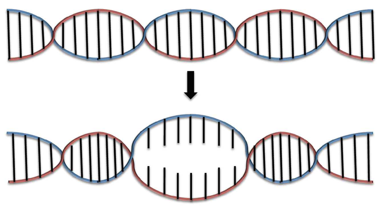 The non-covalent interactions between antiparallel strands in DNA can be broken in order to "open" the double helix when biologically important mechanisms such as DNA replication, transcription, DNA repair or protein binding are set to occur.[10] The area of partially separated DNA is known as the denaturation bubble, which can be more specifically defined as the opening of a DNA double helix through the coordinated separation of base pairs.[10]
The first model that attempted to describe the thermodynamics of the denaturation bubble was introduced in 1966 and called the Poland-Scheraga Model. This model describes the denaturation of DNA strands as a function of temperature. As the temperature increases, the hydrogen bonds between the Watson and Crick base pairs are increasingly disturbed and "denatured loops" begin to form.[11] However, the Poland-Scheraga Model is now considered elementary because it fails to account for the confounding implications of DNA sequence, chemical composition, stiffness and torsion.[12]
Recent thermodynamic studies have inferred that the lifetime of a singular denaturation bubble ranges from 1 microsecond to 1 millisecond.[13] This information is based on established timescales of DNA replication and transcription.[13] Currently, biophysical and biochemical research studies are being performed to more fully elucidate the thermodynamic details of the denaturation bubble.[13]
Denaturation due to Chemical Agents

With polymerase chain reaction (PCR) being among the most popular contexts in which DNA denaturation is desired, heating is the most frequent method of denaturation.[14] Other than denaturation by heat, nucleic acids can undergo the denaturation process through various chemical agents such as formamide, guanidine, sodium salicylate, dimethyl sulfoxide (DMSO), propylene glycol, and urea.[15] These chemical denaturing agents lower the melting temperature (Tm) by competing for hydrogen bond donors and acceptors with pre-existing nitrogenous base pairs. Some agents are even able to induce denaturation at room temperature. For example, alkaline agents (e.g. NaOH) have been shown to denature DNA by changing pH and removing hydrogen-bond contributing protons.[14] These denaturants have been employed to make Denaturing Gradient Gel Electrophoresis gel (DGGE), which promotes denaturation of nucleic acids in order to eliminate the influence of nucleic acid shape on their electrophoretic mobility.[16]
Chemical Denaturation as an Alternative
The optical activity (absorption and scattering of light) and hydrodynamic properties (translational diffusion, sedimentation coefficients, and rotational correlation times) of formamide denatured nucleic acids are similar to those of heat-denatured nucleic acids.[15][17][18] Therefore, depending on the desired effect, chemically denaturing DNA can provide a gentler procedure for denaturing nucleic acids than denaturation induced by heat. Studies comparing different denaturation methods such as heating, beads mill of different bead sizes, probe sonification, and chemical denaturation show that chemical denaturation can provide quicker denaturation compared to the other physical denaturation methods described.[14] Particularly in cases where rapid renaturation is desired, chemical denaturation agents can provide an ideal alternative to heating. For example, DNA strands denatured with alkaline agents such as NaOH denatures as soon as phosphate buffer is added.[14]
Denaturation due to Air
Small, electronegative molecules such as nitrogen and oxygen, which are the primary gases in air, significantly impact the ability of surrounding molecules to participate in hydrogen bonding.[19] These molecules compete with surrounding hydrogen bond acceptors for hydrogen bond donors, therefore acting as "hydrogen bond breakers" and weakening interactions between surrounding molecules in the environment.[19] Antiparellel strands in DNA double helices are non-covalently bound by hydrogen bonding between Watson and Crick base pairs;[20] nitrogen and oxygen therefore maintain the potential to weaken the integrity of DNA when exposed to air.[21] As a result, DNA strands exposed to air require less force to separate and exemplify lower melting temperatures.[21]
Applications
Many laboratory techniques rely on the ability of nucleic acid strands to separate. By understanding the properties of nucleic acid denaturation, the following methods were created:
Denaturants
Protein Denaturants
Acids
Acidic protein denaturants include:
- Acetic acid[22]
- Trichloroacetic acid 12% in water
- Sulfosalicylic acid
Bases
Bases work similarly to acids in denaturation. They include:
Solvents
Most organic solvents are denaturing, including:
Cross-linking reagents
Cross-linking agents for proteins include:
Chaotropic agents
Chaotropic agents include:
- Urea 6 – 8 mol/l
- Guanidinium chloride 6 mol/l
- Lithium perchlorate 4.5 mol/l
Disulfide bond reducers
Agents that break disulfide bonds by reduction include:
- 2-Mercaptoethanol
- Dithiothreitol
- TCEP (tris(2-carboxyethyl)phosphine)
Other
- Mechanical agitation
- Picric acid
- Radiation
- Temperature[23]
Nucleic Acid Denaturants
Chemical
Acidic nucleic acid denaturants include:
- Acetic acid
- HCl
Basic nucleic acid denaturants include:
- NaOH
Other nucleic acid denaturants include:
Physical
- Thermal denaturation
- Beads mill
- Probe sonification
- Radiation
See also
References
- ↑ Alan D. MacNaught; Andrew R. Wilkinson, eds. (1997). Compendium of Chemical Terminology: IUPAC Recommendations (the "Gold Book"). Blackwell Science. ISBN 0865426848.
- ↑ "Terminology for biorelated polymers and applications (IUPAC Recommendations 2012)" (PDF). Pure and Applied Chemistry. 84 (2): 377–410. 2012. doi:10.1351/PAC-REC-10-12-04.
- ↑ Mosby's Medical Dictionary (8th ed.). Elsevier. 2009. Retrieved September 2013. Check date values in:
|access-date=(help) - ↑ Samson, Andre L.; Ho, Bosco; Au, Amanda E.; Schoenwaelder, Simone M.; Smyth, Mark J.; Bottomley, Stephen P.; Kleifeld, Oded; Medcalf, Robert L. (2016-11-01). "Physicochemical properties that control protein aggregation also determine whether a protein is retained or released from necrotic cells". Open Biology. 6 (11). doi:10.1098/rsob.160098. ISSN 2046-2441. PMID 27810968.
- ↑ Samson, Andre L.; Knaupp, Anja S.; Sashindranath, Maithili; Borg, Rachael J.; Au, Amanda E.-L.; Cops, Elisa J.; Saunders, Helen M.; Cody, Stephen H.; McLean, Catriona A. (2012-10-25). "Nucleocytoplasmic coagulation: an injury-induced aggregation event that disulfide crosslinks proteins and facilitates their removal by plasmin". Cell Reports. 2 (4): 889–901. doi:10.1016/j.celrep.2012.08.026. ISSN 2211-1247. PMID 23041318.
- ↑ "Ceviche: the new sushi," The Times.
- ↑ Charles Tanford (1968), "Protein denaturation" (PDF), Advances in Protein Chemistry, 23: 121–282, doi:10.1016/S0065-3233(08)60401-5, PMID 4882248
- ↑ Campbell, N. A.; Reece, J.B.; Meyers, N.; Urry, L. A.; Cain, M.L.; Wasserman, S.A.; Minorsky, P.V.; Jackson, R.B. (2009), Biology (8th, Australian version ed.), Sydney: Pearson Education Australia
- ↑ Anfinsen CB. (1973), "Principles that govern the folding of protein chains", Science, 181 (4096): 223–30, doi:10.1126/science.181.4096.223, PMID 4124164
- 1 2 Sicard, François; Destainville, Nicolas; Manghi, Manoel (21 January 2015). "DNA denaturation bubbles: Free-energy landscape and nucleation/closure rates". The Journal of Chemical Physics. 142 (3): 034903. doi:10.1063/1.4905668.
- ↑ Lieu, Simon. "The Poland-Scheraga Model." (2015): 0-5. Massachusetts Institute of Technology, 14 May 2015. Web. 25 Oct. 2016.
- ↑ Richard, C., and A. J. Guttmann. "Poland–Scheraga Models and the DNA Denaturation Transition." Journal of Statistical Physics 115.3/4 (2004): 925-47. Web.
- 1 2 3 Altan-Bonnet, Grégoire; Libchaber, Albert; Krichevsky, Oleg (1 April 2003). "Bubble Dynamics in Double-Stranded DNA". Physical Review Letters. 90 (13). doi:10.1103/physrevlett.90.138101.
- 1 2 3 4 Wang, X (2014). "Characterization of denaturation and renaturation of DNA for DNA hybridization". Environmental Health and Toxicology Environ Health Toxicol. 29. doi:10.5620/eht.2014.29.e2014007.
- 1 2 Marmur, J (1961). "Denaturation of deoxyribonucleic acid by formamide". Biochimica Et Biophysica Acta. 51 (1): 91013–7.
- ↑ "Denaturing Polyacrylamide Gel Electrophoresis of DNA & RNA". Electrophoresis. National Diagnostics. Retrieved 13 October 2016.
- ↑ Tinoco, I; Bustamante, C; Maestre, M (1980). "The Optical Activity of Nucleic Acids and their Aggregates". Annual Review of Biophysics and Bioengineering. 9 (1): 107–141. doi:10.1146/annurev.bb.09.060180.000543.
- ↑ Fernandes, M (2002). "Calculation of hydrodynamic properties of small nucleic acids from their atomic structure". Nucleic Acids Research. 30 (8): 1782–8. doi:10.1093/nar/30.8.1782.
- 1 2 Mathers, T. L.; Schoeffler, G.; McGlynn, S. P. (July 1985). "The effects of selected gases upon ethanol: hydrogen bond breaking by O and N". Canadian Journal of Chemistry. 63 (7): 1864–1869. doi:10.1139/v85-309.
- ↑ Cox, David L. Nelson, Michael M. (2008). Lehninger principles of biochemistry (5th ed. ed.). New York: W.H. Freeman. ISBN 9780716771081.
- 1 2 Mathers, T. L.; Schoeffler, G.; McGlynn, S. P. (1982). "Hydrogen-bond breaking by O/sub 2/ and N/sub 2/. II. Melting curves of DNA". doi:10.2172/5693881.
- ↑ López-Alonso JP, Bruix M, Font J, Ribó M, Vilanova M, Jiménez MA, Santoro J, González C, Laurents DV (2010), "NMR spectroscopy reveals that RNase A is chiefly denatured in 40% acetic acid: implications for oligomer formation by 3D domain swapping", J. Am. Chem. Soc., 132 (5): 1621–30, doi:10.1021/ja9081638, PMID 20085318
- ↑ Jaremko, M.; Jaremko Ł; Kim HY; Cho MK; Schwieters CD; Giller K; Becker S; Zweckstetter M. (April 2013). "Cold denaturation of a protein dimer monitored at atomic resolution". Nat. Chem. Biol. 9 (4): 264–70. doi:10.1038/nchembio.1181. PMID 23396077.