DnaA
| Chromosomal replication initiator protein dnaA | |
|---|---|
| Identifiers | |
| Organism | |
| Symbol | DnaA |
| Entrez | 948217 |
| RefSeq (Prot) | NP_418157.1 |
| UniProt | P03004 |
| Other data | |
| Chromosome | genome: 3.88 - 3.88 Mb |
| Bac_DnaA_C | |||||||||
|---|---|---|---|---|---|---|---|---|---|
 crystal structure of dnaa domainiv complexed with dnaabox dna | |||||||||
| Identifiers | |||||||||
| Symbol | Bac_DnaA_C | ||||||||
| Pfam | PF08299 | ||||||||
| Pfam clan | CL0123 | ||||||||
| InterPro | IPR013159 | ||||||||
| SCOP | 1j1v | ||||||||
| SUPERFAMILY | 1j1v | ||||||||
| |||||||||
| Bac_DnaA | |||||||||
|---|---|---|---|---|---|---|---|---|---|
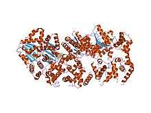 structure of amppcp-bound dnaa from aquifex aeolicus | |||||||||
| Identifiers | |||||||||
| Symbol | Bac_DnaA | ||||||||
| Pfam | PF00308 | ||||||||
| Pfam clan | CL0023 | ||||||||
| InterPro | IPR013317 | ||||||||
| PROSITE | PDOC00771 | ||||||||
| SCOP | 1j1v | ||||||||
| SUPERFAMILY | 1j1v | ||||||||
| |||||||||
DnaA is a protein that activates initiation of DNA replication in bacteria.[1] It is a replication initiation factor which promotes the unwinding of DNA at oriC.[1] The onset of the initiation phase of DNA replication is determined by the concentration of DnaA.[1] DnaA accumulates during growth and then triggers the initiation of replication.[1] Replication begins with active DnaA binding to 9-mer (9-bp) repeats upstream of oriC.[1] Binding of DnaA leads to strand separation at the 13-mer repeats.[1] This binding causes the DNA to loop in preparation for melting open by the helicase DnaB.[1]
Function
The active form DnaA is bound to ATP.[1] Immediately after a cell has divided, the level of active DnaA within the cell is low.[1] Although the active form of DnaA requires ATP, the formation of the oriC/DnaA complex and subsequent DNA unwinding does not require ATP hydrolysis.[2]
The oriC site in E. coli has three AT rich 13 base pair regions (DUEs) followed by four 9 bp regions with the sequence TTAT(C or A)CA(C or A)A.[3] Around 10 DnaA molecules bind to the 9 bp regions, which wrap around the proteins causing the DNA at the AT-rich region to unwind. There are 8 DnaA binding sites within oriC, to which DnaA binds with differential affinity. When DNA replication is about to commence, DnaA occupies all of the high and low affinity binding sites. The denatured AT-rich region allows for the recruitment of DnaB (helicase), which complexes with DnaC (helicase loader). DnaC helps the helicase to bind to and to properly accommodate the ssDNA at the 13 bp region; this is accomplished by ATP hydrolysis, after which DnaC is released. Single-strand binding proteins (SSBs) stabilize the single DNA strands in order to maintain the replication bubble. DnaB is a 5'→3' helicase, so it travels on the lagging strand. It associates with DnaG (a primase) to form the only primer for the leading strand and to add RNA primers on the lagging strand. The interaction between DnaG and DnaB is necessary to control the longitude of Okazaki fragments on the lagging strand. DNA polymerase III is then able to start DNA replication.
DnaA contains two conserved regions: the first is located in the central part of the protein and corresponds to the ATP-binding domain, the second is located in the C-terminal half and is involved in DNA-binding.[4]
See Also
References
- 1 2 3 4 5 6 7 8 9 Foster JB, Slonczewski J (2009). Microbiology: an evolving science. New York: W.W. Norton & Co. ISBN 0-393-97857-5.
- ↑ Leonard AC, Grimwade JE (December 2010). "Regulating DnaA complex assembly: it is time to fill the gaps". Curr. Opin. Microbiol. 13 (6): 766–72. doi:10.1016/j.mib.2010.10.001. PMC 3005629
 . PMID 21035377.
 - ↑ Fuller RS, Funnell BE, Kornberg A (October 1984). "The dnaA protein complex with the E. coli chromosomal replication origin (oriC) and other DNA sites". Cell. 38 (3): 889–900. doi:10.1016/0092-8674(84)90284-8. PMID 6091903.
- ↑ Roth A, Messer W (May 1995). "The DNA binding domain of the initiator protein DnaA". EMBO J. 14 (9): 2106–11. PMC 398312. PMID 7744016.
Further reading
External links
- DnaA protein, Bacteria at the US National Library of Medicine Medical Subject Headings (MeSH)
This article incorporates text from the public domain Pfam and InterPro IPR013159
This article incorporates text from the public domain Pfam and InterPro IPR013317