Habenular nuclei
| Habenular nuclei | |
|---|---|
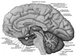 Mesal aspect of a brain sectioned in the median sagittal plane. The habenular nuclei are not labeled directly, but after expanding, look to region with 'habenular commissure', 'pineal body', and 'posterior commissure' | |
| Details | |
| Identifiers | |
| Latin | nucleus habenularis lateralis, nucleus habenularis medialis |
| TA |
A14.1.08.503 A14.1.08.504 |
| FMA | 62372 |
The Habenular nuclei, latin for "little rein," acts as a regulator of key central nervous system neurotransmitters, connecting the forebrain and midbrain within the epithalamus.[1][2][3] Although predominantly studied for its demonstration of asymmetrical brain development and function, in recent years many scientists have begun to examine the Habenular nuclei's role in motivation and behavior as it relates to an understanding of the physiology of addiction.
Anatomy and connectivity
The habenular nuclei comprise a small group of nuclei that are part of the epithalamus of the diencephalon, and is located just above the thalamus and is divided into two asymmetric halves, the medial habenula (MHb) and the lateral habenula (LHb) that regulate monamines, such as dopamine and serotonin.[4][5] Information from the posterior septum and a portion of Broca's area feeds into the MHb, and a region of the hypothalamus, nucleus accumbens, ventral pallidum, globus pallidus, and a portion of Broca's area all feed into the LHb.[2] As a whole, this complexly interconnected region is part of the dorsal diencephalic conduction (DDC) system, responsible for relaying information from the limbic system to the midbrain, hindbrain, and medial forebrain.[6][7]
Habenula Nuclear divisions:
- lateral habenular nucleus (hier-278 at NeuroNames, )
- medial habenular nucleus (hier-279 at NeuroNames, )
The pineal gland is attached to the brain in this region.
Nerve impulses from the habenular nuclei are transmitted to the septal nuclei via the stria medullaris, which is found on the medial surface of the thalamus.
Motivation and addiction
Recent exploration of the Habenular nuclei has begun to associate the structure with an organism's current mood, feeling of motivation, and reward recognition.[8] Previously, the LHb has been identified as an "anti-reward" signal, but recent research suggests that the LHb helps identify preference, helping the brain to discriminate between potential actions and subsequent motivation decisions.[9] In a study using a Pavlovian conditioning model, results showed an increase in the habenula response.[10] This increase coincided with conditioned stimuli associated with more aversive punishments (ie. electric shock).[10] Therefore, researchers speculate that inhibition or damage to the LHb resulting in a failure to process such information may lead to random motivation behavior.[9][10]
LHb is especially important in understanding the reward and motivation relationship as it relates to addictive behaviors.[8] The LHb inhibits dopaminergic neurons, decreasing the release of dopamine.[11] It was determined by several animal studies that receiving a reward coincided with elevated dopamine levels, but once the learned association was learned by the animal, dopamine levels remain elevated, only decreasing when the reward is removed.[1][5][8][11] Therefore, dopamine levels only increase with unpredicted rewards and with a "negative prediction error".[1] Moreover, it was determined that removal of an anticipated award activated LHb, inhibited dopamine levels.[1] This finding helps explain why addictive drugs are associated with elevated dopamine levels.[1]
Nicotine and nAChRs
According to the National Institute on Drug Abuse, 1 in 5 deaths in the United States is caused by tobacco use.[12] Nicotine is the addictive drug found in most tobacco products and is easily absorbed by the bloodstream of the body.[12] Despite common misconceptions regarding the relaxing effects of tobacco and nicotine use, behavioral testing in animals has demonstrated nicotine to have an anxiogenic effect.[13] Nicotinic acetylcholine receptors, or nAChRs, have been identified as the primary site for nicotine activity and regulate consequent cellular polarization.[14] nAChRs are made up a number of α and β subunits and are found in both the LHb and MHb where research suggests they may play a key role in addiction and withdrawal behaviors.[14][15]
References
- 1 2 3 4 5 Velasquez, Kenia Marisela; Molfese, David Lucas; Salas, Ramiro (2014-01-01). "The role of the habenula in drug addiction". Frontiers in Human Neuroscience. 8: 174. doi:10.3389/fnhum.2014.00174. PMC 3975120
 . PMID 24734015.
 - 1 2 Aizawa, Hidenori (2012-10-20). "Habenula and the asymmetric development of the vertebrate brain". Anatomical Science International. 88 (1): 1–9. doi:10.1007/s12565-012-0158-6. ISSN 1447-6959.
- ↑ Aizawa, Hidenori; Amo, Ryunosuke; Okamoto, Hitoshi (2011-01-01). "Phylogeny and ontogeny of the habenular structure". Neurogenesis. 5: 138. doi:10.3389/fnins.2011.00138. PMC 3244072. PMID 22203792.
- ↑ Stephenson-Jones, Marcus (2011). "Evolutionary conservation of the habenular nuclei and their circuitry controlling the dopamine and 5-hydroxytryptophan ( 5-HT ) systems". Proceedings of the National Academy of Sciences of the United States of America.
- 1 2 Boulos, Laura-Joy; Darcq, Emmanuel; Kieffer, Brigitte Lina. "Translating the Habenula—From Rodents to Humans". Biological Psychiatry. doi:10.1016/j.biopsych.2016.06.003.
- ↑ Beretta, Carlo Antonio; Dross, Nicolas; Gutierrez-Triana, Jose Arturo; Ryu, Soojin; Carl, Matthias (2012-01-01). "Habenula circuit development: past, present, and future". Neurogenesis. 6: 51. doi:10.3389/fnins.2012.00051. PMC 3332237. PMID 22536170.
- ↑ Bianco, Isaac H.; Wilson, Stephen W. (2009-04-12). "The habenular nuclei: a conserved asymmetric relay station in the vertebrate brain". Philosophical Transactions of the Royal Society of London B: Biological Sciences. 364 (1519): 1005–1020. doi:10.1098/rstb.2008.0213. ISSN 0962-8436. PMC 2666075. PMID 19064356.
- 1 2 3 Fakhoury, Marc; López, Domínguez. "The Role of Habenula in Motivation and Reward". Advances in Neuroscience.
- 1 2 Stopper, Colin M; Floresco, Stan B (24 November 2013). "What's better for me? Fundamental role for lateral habenula in promoting subjective decision biases". Nature Neuroscience. 17 (1): 33–35. doi:10.1038/nn.3587.
- 1 2 3 Lawson, Rebecca P.; Seymour, Ben; Loh, Eleanor; Lutti, Antoine; Dolan, Raymond J.; Dayan, Peter; Weiskopf, Nikolaus; Roiser, Jonathan P. (2014-08-12). "The habenula encodes negative motivational value associated with primary punishment in humans". Proceedings of the National Academy of Sciences. 111 (32): 11858–11863. doi:10.1073/pnas.1323586111. ISSN 0027-8424. PMC 4136587. PMID 25071182.
- 1 2 Hikosaka, Okihide; Sesack, Susan R.; Lecourtier, Lucas; Shepard, Paul D. (2008-11-12). "Habenula: Crossroad between the Basal Ganglia and the Limbic System". Journal of Neuroscience. 28 (46): 11825–11829. doi:10.1523/jneurosci.3463-08.2008.
- 1 2 Abuse, National Institute on Drug (2014-12-16). "Tobacco/Nicotine". Retrieved 2016-11-22.
- ↑ Casarrubea, Maurizio; Davies, Caitlin; Faulisi, Fabiana; Pierucci, Massimo; Colangeli, Roberto; Partridge, Lucy; Chambers, Stephanie; Cassar, Daniel; Valentino, Mario (2015-01-01). "Acute nicotine induces anxiety and disrupts temporal pattern organization of rat exploratory behavior in hole-board: a potential role for the lateral habenula". Frontiers in Cellular Neuroscience: 197. doi:10.3389/fncel.2015.00197. PMC 4450172. PMID 26082682.
- 1 2 Zuo, Wanhong; Xiao, Cheng; Gao, Ming; Hopf, F. Woodward; Krnjević, Krešimir; McIntosh, J. Michael; Fu, Rao; Wu, Jie; Bekker, Alex (2016-09-06). "Nicotine regulates activity of lateral habenula neurons via presynaptic and postsynaptic mechanisms". Scientific Reports. 6. doi:10.1038/srep32937. ISSN 2045-2322. PMC 5011770. PMID 27596561.
- ↑ Dao, Dang Q.; Perez, Erika E.; Teng, Yanfen; Dani, John A.; De Biasi, Mariella (2014-03-19). "Nicotine Enhances Excitability of Medial Habenular Neurons via Facilitation of Neurokinin Signaling". Journal of Neuroscience. 34 (12): 4273–4284. doi:10.1523/jneurosci.2736-13.2014.