Hemocyanin
| Hemocyanin, copper containing domain | |||||||||
|---|---|---|---|---|---|---|---|---|---|
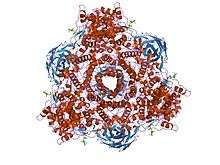
 Single Oxygenated Functional Unit from the hemocyanin of an octopus | |||||||||
| Identifiers | |||||||||
| Symbol | Hemocyanin_M | ||||||||
| Pfam | PF00372 | ||||||||
| InterPro | IPR000896 | ||||||||
| PROSITE | PDOC00184 | ||||||||
| SCOP | 1lla | ||||||||
| SUPERFAMILY | 1lla | ||||||||
| |||||||||
| Hemocyanin, all-alpha domain | |||||||||
|---|---|---|---|---|---|---|---|---|---|
 Crystal structure of hexameric haemocyanin from Panulirus interruptus refined at 3.2 angstroms resolution | |||||||||
| Identifiers | |||||||||
| Symbol | Hemocyanin_N | ||||||||
| Pfam | PF03722 | ||||||||
| InterPro | IPR005204 | ||||||||
| PROSITE | PDOC00184 | ||||||||
| SCOP | 1lla | ||||||||
| SUPERFAMILY | 1lla | ||||||||
| |||||||||
| Hemocyanin, ig-like domain | |||||||||
|---|---|---|---|---|---|---|---|---|---|
 crystallographic analysis of oxygenated and deoxygenated states of arthropod hemocyanin shows unusual differences | |||||||||
| Identifiers | |||||||||
| Symbol | Hemocyanin_C | ||||||||
| Pfam | PF03723 | ||||||||
| InterPro | IPR005203 | ||||||||
| PROSITE | PDOC00184 | ||||||||
| SCOP | 1lla | ||||||||
| SUPERFAMILY | 1lla | ||||||||
| |||||||||
Hemocyanins (also spelled haemocyanins and abbreviated Hc) are proteins that transport oxygen throughout the bodies of some invertebrate animals. These metalloproteins contain two copper atoms that reversibly bind a single oxygen molecule (O2). They are second only to hemoglobin in frequency of use as an oxygen transport molecule. Unlike the hemoglobin in red blood cells found in vertebrates, hemocyanins are not bound to blood cells but are instead suspended directly in the hemolymph. Oxygenation causes a color change between the colorless Cu(I) deoxygenated form and the blue Cu(II) oxygenated form.[1]
Species distribution
Hemocyanins are found only in the Mollusca and Arthropoda: the earliest hemocyanins were found in the snail Helix pomatia (a mollusc) and in the horseshoe crab (an arthropod). They were subsequently found to be common among crustaceans and are utilized by some land arthropods such as the tarantula Eurypelma californicum,[2] the emperor scorpion,[3] and the centipede Scutigera coleoptrata. Also, larval storage proteins in many insects appear to be derived from hemocyanins.
The hemocyanin superfamily
The arthropod hemocyanin superfamily is composed of phenoloxidases, hexamerins, pseudohemocyanins or cryptocyanins, (dipteran) hexamerin receptors.
Phenoloxidase are copper containing tyrosinases. These proteins are involved in the process of sclerotization of arthropod cuticle, in wound healing, and humoral immune defense. Phenoloxidase is synthesized by zymogens and are activated by cleaving a N-terminal peptide.
Hexamerins are storage proteins commonly found in insects. These proteins are synthesized by the larval fat body and are associated with molting cycles or nutritional conditions
Pseudohemocyanin and cryptocyanins genetic sequences are closely related to hemocyanins in crustaceans. These proteins have a similar structure and function, but lack the copper binding sites.
The evolutionary changes within the phylogeny of the hemocyanin superfamily are closely related to the emergence of these different proteins in various species. The understanding of proteins within this superfamily would not be well understood without the extensive studies of hemocyanin in arthropods.[4]
Structure and mechanism
Although the respiratory function of hemocyanin is similar to that of hemoglobin, there are a significant number of differences in its molecular structure and mechanism. Whereas hemoglobin carries its iron atoms in porphyrin rings (heme groups), the copper atoms of hemocyanin are bound as prosthetic groups coordinated by histidine residues. The active site of hemocyanin is composed of a pair of copper(I) cations which are directly coordinated to the protein through the driving force of imidazolic rings of six histidine residues.[5] It has been noted that species using hemocyanin for oxygen transportation include crustaceans living in cold environments with low oxygen pressure. Under these circumstances hemoglobin oxygen transportation is less efficient than hemocyanin oxygen transportation.[6] Nevertheless, there are also terrestrial arthropods using hemocyanin, notably spiders and scorpions, that live in warm climates.
Most hemocyanins bind with oxygen non-cooperatively and are roughly one-fourth as efficient as hemoglobin at transporting oxygen per amount of blood. Hemoglobin binds oxygen cooperatively due to steric conformation changes in the protein complex, which increases hemoglobin's affinity for oxygen when partially oxygenated. In some hemocyanins of horseshoe crabs and some other species of arthropods, cooperative binding is observed, with Hill coefficients of 1.6 - 3.0. Hill coefficients vary depending on species and laboratory measurement settings. Hemoglobin, for comparison, has a Hill coefficient of usually 2.8 - 3.0. In these cases of cooperative binding hemocyanin was arranged in protein sub-complexes of 6 subunits (hexamer) each with one oxygen binding site; binding of oxygen on one unit in the complex would increase the affinity of the neighboring units. Each hexamer complex was arranged together to form a larger complex of dozens of hexamers. In one study, cooperative binding was found to be dependent on hexamers being arranged together in the larger complex, suggesting cooperative binding between hexamers. Hemocyanin oxygen-binding profile is also affected by dissolved salt ion levels and pH.[7]
Hemocyanin is made of many individual subunit proteins, each of which contains two copper atoms and can bind one oxygen molecule (O2). Each subunit weighs about 75 kilodaltons (kDa). Subunits may be arranged in dimers or hexamers depending on species; the dimer or hexamer complex is likewise arranged in chains or clusters with weights exceeding 1500 kDa. The subunits are usually homogeneous, or heterogeneous with two variant subunit types. Because of the large size of hemocyanin, it is usually found free-floating in the blood, unlike hemoglobin.[8]
Hexamers are characteristic of arthropod hemocyanins.[9] A hemocyanin of the tarantula Eurypelma californicum[2] is made up of 4 hexamers or 24 pepide chains. A hemocyanin from the house centipede Scutigera coleoptrata[10] is made up of 6 hexamers or 36 chains. Horseshoe crabs have an 8-hexamer (i. e. 48-chain) hemocyanin. Simple hexamers are found in the spiny lobster Panulirus interruptus and the isopod Bathynomus giganteus.[11] Peptide chains in crustaceans are about 660 amino acid residues long, and in chelicerates they are about 625. In the large complexes there is a variety of variant chains, all about the same length; pure components do not usually self-assemble.
Catalytic activity
Hemocyanin is homologous to the phenol oxidases (e.g. tyrosinase) since both enzymes share type 3 Cu active site coordination.[12] In both cases inactive proenzymes such as hemocyanin, tyrosinase, and catcholoxidase must be activated first. This is done by removing the amino acid that blocks the entrance channel to the active site when the proenzyme is not active. There is currently no other known modifications necessary to activate the proenzyme and enable catalytic activity. Conformational differences determine the type of catalytic activity that the hemocyanin is able to perform.[13] Hemocyanin also exhibits phenol oxidase activity, but with slowed kinetics from greater steric bulk at the active site. Partial denaturation actually improves hemocyanin’s phenol oxidase activity by providing greater access to the active site.[1][12]
Spectral properties


Spectroscopy of oxyhemocyanin shows several salient features:[14]
- Resonance Raman spectroscopy shows symmetric binding
- UV-Vis spectroscopy shows strong absorbances at 350 (20000) and 580 (1000) nm
- OxyHc is EPR-silent indicating the absence of unpaired electrons
- Infrared spectroscopy shows ν(O-O) of 755 cm−1
(1) rules out a mononuclear peroxo complex (2) does not match with the UV-Vis spectra of mononuclear peroxo and Kenneth D. Karlin's dinuclear end-on coordinated trans-peroxo model complexes.[15] (4) shows a considerably weaker O-O bond compared with Karlin's trans-peroxo model.[15]
On the other hand, Nobumasa Kitajima's dinuclear side-on coordinated peroxo model complexes shows ν(O-O) of 741 cm−1 and UV-Vis absorbances at 349 and 551 nm, which agree with the experimental observations for oxyHc.[16] The Cu-Cu separation in the model complex is 3.56 Å, that of oxyhemocyanin is ca. 3.6 Å (deoxyHc: ca. 4.6 Å).[16][17][18]
Anticancer effects
The hemocyanin found in Concholepas concholepas blood has immunotherapeutic effects against bladder cancer in murine models. Researchers in 2006 primed mice with C. concholepas before implantation of bladder tumor (MBT-2) cells. Mice treated with C. concholepas hemocyanin showed antitumor effects: prolonged survival, decreased tumor growth and incidence, and lack of toxic effects and may have a potential use in future immunotherapy for superficial bladder cancer.[19]
Keyhole limpet hemocyanin (KLH) is a known immune stimulant derived from circulating glycoproteins of the marine mollusk Megathura crenulata. KLH has been shown to be a significant treatment against the proliferations of breast cancer, pancreas cancer, and prostate cancer cells when delivered in vitro. The 2003 research study "Keyhole limpet hemocyanin, a novel immune stimulant with promising anticancer activity in Barrett’s esophageal adenocarcinoma" shows that keyhole limpet hemocyanin is also effective in the inhibition of growth of human Barrett’s esophageal cancer through both apoptic and nonapoptic mechanisms of cell death. This research is promising for future cancer curative research.[20]
Case studies: environmental impact on hemocyanin levels
A 2003 study of the effect of culture conditions of blood metabolites and hemocyanin of the white shrimp Litopenaeus vannamei found that the levels of hemocyanin, oxyhemocyanin in particular, are affected by the diet. The study compared oxyhemocyanin levels in the blood of white shrimp housed in an indoor pond with a commercial diet with that of white shrimp housed in an outdoor pond with a more readily available protein source (natural live food) as well. Oxyhemocyanin and blood glucose levels were higher in shrimp housed in outdoor ponds. It was also found that blood metabolite levels tended to be lower in low activity level species, such as crabs, lobsters, and the indoor shrimp when compared to the outdoor shrimp. This correlation is possibly indicative of the morphological and physiological evolution of crustaceans. The levels of these blood proteins and metabolites appear to be dependence on energetic demands and availability of those energy sources.[21]
See also
| Wikimedia Commons has media related to Hemocyanin. |
References
- 1 2 Coates CJ, Nairn J (July 2014). "Diverse immune functions of hemocyanins". Dev. Comp. Immunol. 45 (1): 43–55. doi:10.1016/j.dci.2014.01.021. PMID 24486681.
- 1 2 Voit R, Feldmaier-Fuchs G, Schweikardt T, Decker H, Burmester T (December 2000). "Complete sequence of the 24-mer hemocyanin of the tarantula Eurypelma californicum. Structure and intramolecular evolution of the subunits". J. Biol. Chem. 275 (50): 39339–44. doi:10.1074/jbc.M005442200. PMID 10961996.
- ↑ Jaenicke E, Pairet B, Hartmann H, Decker H (2012). "Crystallization and preliminary analysis of crystals of the 24-meric hemocyanin of the emperor scorpion (Pandinus imperator)". PLoS ONE. 7 (3): e32548. doi:10.1371/journal.pone.0032548. PMC 3293826
 . PMID 22403673. Lay summary – Johannes Gutenberg-Universität Mainz.
 - ↑ Burmester, T. (2001). "Molecular Evolution of the Arthropod Hemocyanin Superfamily". Mol. Biol. Evol.. 18 (2): 184–195. doi:10.1093/oxfordjournals.molbev.a003792.
- ↑ Rannulu, N. S.; Rodgers, M. T. (2005). "Solvation of copper ions by imidazole: Structure and sequential binding energies of Cu+ (imidazole)x, x = 1-4. Competition between ion solvation and hydrogen bonding". Phys. Chem. Chem. Phys. 7: 1014–1025–23. doi:10.1039/b418141g.
- ↑ Strobel A, Hu MY, Gutowska MA, Lieb B, Lucassen M, Melzner F, Pörtner HO, Mark FC (December 2012). "Influence of temperature, hypercapnia, and development on the relative expression of different hemocyanin isoforms in the common cuttlefish Sepia officinalis". J Exp Zool a Ecol Genet Physiol. 317 (8): 511–23. doi:10.1002/jez.1743. PMID 22791630.
- ↑ Perton FG, Beintema JJ, Decker H (May 1997). "Influence of antibody binding on oxygen binding behavior of Panulirus interruptus hemocyanin". FEBS Lett. 408 (2): 124–6. doi:10.1016/S0014-5793(97)00269-X. PMID 9187351.
- ↑ Waxman L (May 1975). "The structure of arthropod and mollusc hemocyanins". J. Biol. Chem. 250 (10): 3796–806. PMID 1126935.
- ↑ (van Holde & Miller 1995, p. 9)
- ↑ Kusche K, Hembach A, Hagner-Holler S, Gebauer W, Burmester T (July 2003). "Complete subunit sequences, structure and evolution of the 6 x 6-mer hemocyanin from the common house centipede, Scutigera coleoptrata". Eur. J. Biochem. 270 (13): 2860–8. doi:10.1046/j.1432-1033.2003.03664.x. PMID 12823556.
- ↑ (van Holde & Miller 1995, p. 8)
- 1 2 Decker H, Tuczek F (August 2000). "Tyrosinase/catecholoxidase activity of hemocyanins: structural basis and molecular mechanism". Trends Biochem. Sci. 25 (8): 392–7. doi:10.1016/S0968-0004(00)01602-9. PMID 10916160.
- ↑ Decker, H.; Schweikardt, T.; Nillius, D.; Salzbrunn, U.; Jaenicke, E.; Tuczek, F. (2007). "Similar enzyme activation and catalysis in hemocyanins and tyrosinases.". 398 (1-2): 183–191. doi:10.1016/j.gene.2007.02.051.
- ↑ Freedman TB, Loehr JS, Loehr TM (1976). "A resonance Raman study of the copper protein, hemocyanin. New evidence for the structure of the oxygen-binding site". J. Am. Chem. Soc. 98 (10): 2809–2815. doi:10.1021/ja00426a023.
- 1 2 Karlin KD, Cruse RW, Gultneh Y, Farooq A, Hayes JC, Zubieta J (1987). "Dioxygen-copper reactivity. Reversible binding of O2 and CO to a phenoxo-bridged dicopper(I) complex". J. Am. Chem. Soc. 109 (9): 2668–2679. doi:10.1021/ja00243a019.
- 1 2 Kitajima N, Fujisawa K, Fujimoto C, Morooka Y, Hashimoto S, Kitagawa T, Toriumi K, Tatsumi K, Nakamura A (1992). "A new model for dioxygen binding in hemocyanin. Synthesis, characterization, and molecular structure of the μ-η2:η2 peroxo dinuclear copper(II) complexes, [Cu(HB(3,5-R2pz)3)]2(O2) (R = isopropyl and Ph)". J. Am. Chem. Soc. 114 (4): 1277–1291. doi:10.1021/ja00030a025.
- ↑ Gaykema WPJ; Hol WGJ; Vereijken JM; Soeter NM; Bak HJ; Beintema JJ (1984). "3.2 Å structure of the copper-containing, oxygen-carrying protein Panulirus interruptus haemocyanin". Nature. 309: 23–29. doi:10.1038/309023a0.
- ↑ Kodera M, Katayama K, Tachi Y, Kano K, Hirota S, Fujinami S, Suzuki M (1999). "Crystal Structure and Reversible O2-Binding of a Room Temperature Stable μ-η2:η2-Peroxodicopper(II) Complex of a Sterically Hindered Hexapyridine Dinucleating Ligand". J. Am. Chem. Soc. 121 (47): 11006–11007. doi:10.1021/ja992295q.
- ↑ Atala A (December 2006). "This Month in Investigative Urology". The Journal of Urology. 176 (6): 2335–2336. doi:10.1016/j.juro.2006.09.002.
- ↑ McFadden, D. W.; Riggs, D. R.; Jackson, B.J.; Vona-Davis, L. (2003). "Keyhole limpet hemocyanin, a novel immune stimulant with promising anticancer activity in Barrett's esophageal adenocarcinoma.". 186 (5): 552–555. doi:10.1016/j.amjsurg.2003.08.002.
- ↑ Pascual, C.; Gaxiola, G.; Rosas, C. (2003). "Blood metabolites and hemocyanin of the white shrimp, Litopenaeus vannamei the effect of culture conditions and a comparison with other crustacean species.". Marine Biology . 142 (4): 735–745.
Further reading
- Rehm P, Pick C, Borner J, Markl J, Burmester T (2012). "The diversity and evolution of chelicerate hemocyanins". BMC Evol. Biol. 12: 19. doi:10.1186/1471-2148-12-19. PMC 3306762. PMID 22333134.
- Ali SA, Abbasi A (2011). Scorpion Hemocyanin: The blue blood. Saarbrücken: VDM Verlag Dr. Müller. p. 160. ISBN 978-3639337259.