Intravenous therapy
| Intravenous therapy | |
|---|---|
| Intervention | |
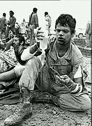
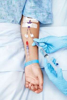 Infusion therapy: A person receiving medication via intravenous therapy. | |
| Synonyms | IV therapy, iv therapy |
Intravenous therapy is the infusion of liquid substances directly into a vein. Intravenous simply means "within vein". Therapies administered intravenously are often included in the designation of specialty drugs. Intravenous infusions are commonly referred to as drips because many systems of administration employ a drip chamber, which prevents air from entering the blood stream (air embolism), and allows an estimation of flow rate.
Intravenous therapy may be used to correct electrolyte imbalances, to deliver medications, for blood transfusion or as fluid replacement to correct, for example, dehydration. Intravenous therapy could also be used for chemotherapy.
Compared with other routes of administration, the intravenous route is the fastest way to deliver fluids and medications throughout the body. The bioavailability of the medication is 100% in IV therapy.
Medical uses
Substances that may be infused intravenously include volume expanders, blood-based products, blood substitutes, medications and nutrition.
Volume expanders
There are two main types of volume expander: crystalloids and colloids. Crystalloids are aqueous solutions of mineral salts or other water-soluble molecules. Colloids contain larger insoluble molecules, such as gelatin. Blood is a colloid.
- The most commonly used crystalloid fluid is normal saline, a solution of sodium chloride at 0.9% concentration, which is close to the concentration in the blood (isotonic). Lactated Ringer's (also known as Ringer's lactate) and the closely related Ringer's acetate, are mildly hypotonic solutions often used in those who have significant burns.
- Colloids preserve a high colloid osmotic pressure in the blood, while, on the other hand, this parameter is decreased by crystalloids due to hemodilution.[1] There does not appear to be a benefit of using colloids over crystalloids.[1] Crystalloids generally are much cheaper than colloids.[1](blood, albumin, plasma, etc.)
- Volume expanders may either be isotonic, hypotonic, or hypertonic. Hypotonic fluids are not generally recommended in children due to increased risk of adverse effects.[2]
The best way to determine if a person will benefit from fluids is by doing a passive leg raise followed by measuring the output from the heart.[3]
Blood-based products
A blood product (or blood-based product) is any component of blood which is collected from a donor for use in a blood transfusion. Blood transfusions can be life-saving in some situations, such as massive blood loss due to trauma, or can be used to replace blood lost during surgery. Blood transfusions may also be used to treat a severe anaemia or thrombocytopenia caused by a blood disease. People with hemophilia usually need a replacement of clotting factor, which is a small part of whole blood. People with sickle-cell disease may require frequent blood transfusions. Early blood transfusions consisted of whole blood, but modern medical practice commonly uses only components of the blood, such as fresh frozen plasma or cryoprecipitate.
Blood substitutes
Blood substitutes (also called artificial blood or blood surrogates) are artificial substances aiming to provide an alternative to blood-based products acquired from donors.
The main blood substitutes used today are volume expanders such as crystalloids and colloids mentioned above. Also, oxygen-carrying substitutes are emerging.
Buffer solutions
Buffer solutions are used to correct acidosis or alkalosis. Lactated Ringer's solution also has some buffering effect. A solution more specifically used for buffering purpose is intravenous sodium bicarbonate.
Other medications
Medications may be mixed into the fluids mentioned above. Certain types of medications can only be given intravenously, such as when there is insufficient uptake by other routes of administration such as enterally. Examples include intravenous immunoglobulin and propofol.
Other
- Parenteral nutrition is feeding a person intravenously, bypassing the usual process of eating and digestion. The person receives nutritional formulas containing salts, glucose, amino acids, lipids and added vitamins.
- Drug injection used for recreational substances usually enters by the intravenous route.
Adverse effects
Pain
An injection inherently causes pain and is medically invasive. In cases in which a choice between intravenous therapy and oral treatment may be made to achieve the same outcome, such as in the case of mild or moderate dehydration treatment (assuming oral rehydration therapy is an option), then one should avoid using intravenous therapy in place of the less invasive oral option.[4] Children in emergency departments being treated for dehydration in particular have better outcomes with oral treatment because it does not cause the pain or risk the complications of an injection.[4]
Cold spray can decrease the pain of putting in an IV.[5]
Infection
Any break in the skin carries a risk of infection. Although IV insertion is an aseptic procedure, skin-dwelling organisms such as Coagulase-negative staphylococcus or Candida albicans may enter through the insertion site around the catheter, or bacteria may be accidentally introduced inside the catheter from contaminated equipment. Moisture introduced to unprotected IV sites through washing or bathing substantially increases the infection risks.
Infection of IV sites is usually local, causing easily visible swelling, redness, and fever. If bacteria do not remain in one area but spread through the bloodstream, the infection is called septicemia and can be rapid and life-threatening. An infected central IV poses a higher risk of septicemia, as it can deliver bacteria directly into the central circulation.
Phlebitis
Phlebitis is inflammation of a vein that may be caused by infection, the mere presence of a foreign body (the IV catheter) or the fluids or medication being given. Symptoms are warmth, swelling, pain, and redness around the vein. The IV device must be removed and if necessary re-inserted into another extremity.
Due to frequent injections and recurring phlebitis, scar tissue can build up along the vein. The peripheral veins of intravenous drug addicts, and of cancer patients undergoing chemotherapy, become sclerotic and difficult to access over time, sometimes forming a hard, painful “venous cord”.
Infiltration / extravasation
Infiltration occurs when an IV fluid or medication accidentally enters the surrounding tissue rather than the vein. It may occur when the vein itself ruptures (the elderly are particularly prone to fragile veins due to a paucity of supporting tissues), when the vein is damaged during insertion of the intravascular access device, when the device is not sited correctly or when the entry point of the device into the vein becomes the path of least resistance (e.g. if a cannula is in a vein for some time, the vein may scar and close and the only way for fluid to leave is along the outside of the cannula where it enters the vein). Infiltration is an inadvertent administration of a nonvesicant solution/drug into the tissue, which happens so often when the tourniquet isn't removed in a timely fashion. Infiltration is characterized by coolness and pallor to the skin as well as localized swelling or edema. It is treated by removing the intravenous access device and elevating the affected limb so that the collected fluids can drain away. Sometimes injections of hyaluronidase can be used to speed the dispersal of the fluid/drug. Infiltration is one of the most common adverse effects of IV therapy[6] and is usually not serious unless the infiltrated fluid is a medication damaging to the surrounding tissue, most commonly a vesicant or chemotherapeutic agent, in which case it is called extravasation and extensive necrosis can occur.[7][8]
Fluid overload
This occurs when fluids are given at a higher rate or in a larger volume than the system can absorb or excrete. Possible consequences include hypertension, heart failure, and pulmonary edema.
Hypothermia
The human body is at risk of accidentally induced hypothermia when large amounts of cold fluids are infused. Rapid temperature changes in the heart may precipitate ventricular fibrillation.
Electrolyte imbalance
Administering a too-dilute or too-concentrated solution can disrupt the patient's balance of sodium, potassium, magnesium, and other electrolytes. Hospital patients usually receive blood tests to monitor these levels . it is essential to correct.
Embolism
A blood clot or other solid mass, as well as an air bubble, can be delivered into the circulation through an IV and end up blocking a vessel; this is called embolism. It is nearly impossible to inject air through a peripheral IV at a dangerous rate. The risk is greater with a central IV.
Air bubbles of less than 30 microliters are thought to dissolve into the circulation harmlessly. A larger amount of air, if delivered all at once, can cause life-threatening damage, or, if extremely large (3-8 milliliters per kilogram of body weight), can stop the heart.
One reason veins are preferred over arteries for intravascular administration is because the flow will pass through the lungs before passing through the body. Air bubbles can leave the blood through the lungs. A patient with a right-to-left shunt is vulnerable to embolism from smaller amounts of air. Fatality by air embolism is rare, although this is in part because it is so difficult to diagnose.
Glucose
Intravenous glucose is used in some Asian countries such as Korea as a pick-me-up, for "energy," but is not a part of routine medical care in the United States where a glucose solution is a prescription drug. Asian immigrants to the United States are at risk if they seek intravenous glucose treatment. It may be had at store-front clinics catering to Asian immigrants, but, despite having no more effect than drinking sugared water, poses medical risks such as the possibility of infection. It is commonly called "ringer."[9]
Intravenous access devices
These can be used to obtain blood (e.g. for testing), also known as phlebotomy, as well as for the administration of medication and fluids.
Hypodermic needle
The simplest form of intravenous access is by passing a hollow needle through the skin directly into the vein. This needle can be connected directly to a syringe (used either to withdraw blood or deliver its contents into the bloodstream) or may be connected to a length of tubing and thence whichever collection or infusion system is desired.
The most convenient site is often the arm, especially the veins on the back of the hand, or the median cubital vein at the elbow, but any identifiable vein can be used. Often it is necessary to use a tourniquet which restricts the venous drainage of the limb and makes the vein bulge. Once the needle is in place, it is common to draw back slightly on the syringe to aspirate blood, thus verifying that the needle is really in a vein. The tourniquet should be removed before injecting to prevent extravasation of the medication.
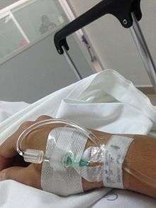
Peripheral cannula


A peripheral cannula is the most common intravenous access method utilized in both hospitals and pre-hospital services. A peripheral IV line (PVC or PIV) consists of a short catheter (a few centimeters long) inserted through the skin into a peripheral vein (any vein not situated in the chest or abdomen). This is usually in the form of a cannula-over-needle device, in which a flexible plastic cannula comes mounted over a metal trocar. Once the tip of the needle and cannula are introduced into the vein via venipuncture, the cannula is advanced inside the vein over the trocar to the appropriate position and secured, the trocar is then withdrawn and discarded. Blood samples may be drawn directly after the initial IV cannula insertion.
Any accessible vein can be used although arm and hand veins are used most commonly, with leg and foot veins used to a much lesser extent. In infants the scalp veins are sometimes used.
The caliber of cannula is commonly indicated in gauge, with 14 being a very large cannula (used in resuscitation settings) and 24-26 the smallest. The most common sizes are 16-gauge (midsize line used for blood donation and transfusion), 18- and 20-gauge (all-purpose line for infusions and blood draws), and 22-gauge (all-purpose pediatric line). 12- and 14-gauge peripheral lines are capable of delivering large volumes of fluid very fast, accounting for their popularity in emergency medicine. These lines are frequently called "large bores" or "trauma lines".
To make the procedure more tolerable for children, medical staff may apply a topical local anaesthetic (such as EMLA or Ametop) to the skin of the chosen venipuncture area about 45 minutes beforehand.
The part of the catheter that remains outside the skin is called the connecting hub; it can be connected to a syringe or an intravenous infusion line, or capped with a heplock or saline lock, a needleless connection filled with a small amount of heparin or saline solution to prevent clotting, between uses of the catheter. Ported cannulae have an injection port on the top that is often used to administer medicine.
In cases of shock, a central venous catheter, a peripherally inserted central catheter (PICC), venous cutdown or intraosseous infusion may be necessary.
Complications
If the cannula is not sited correctly, or the vein is particularly fragile and ruptures, blood may extravasate into the surrounding tissues, this situation is known as a "tissuing" or a "blown vein". Using this cannula to administer medications causes extravasation of the drug which can lead to edema, causing pain and tissue damage, and even necrosis depending on the medication. The person attempting to obtain the access must find a new access site proximal to the "blown" area to prevent extravasation of medications through the damaged vein. For this reason it is advisable to site the first cannula at the most distal appropriate vein.
If a patient needs frequent venous access, the veins may scar and narrow, making any future access extremely difficult or impossible.
A peripheral IV cannot be left in the vein indefinitely, because of the risk of insertion-site infection leading to phlebitis, cellulitis and sepsis. The US Centers for Disease Control and Prevention updated their guidelines and now advise the cannula need to be replaced every 96 hours.[10] This was based on studies organised to identify causes of Methicillin-resistant Staphylococcus aureus (MRSA) infection in hospitals. In the United Kingdom, the UK Department of health published their finding about risk factors associated with increased MRSA infection, now include intravenous cannula, central venous catheters and urinary catheters as the main factors increasing the risk of spreading antibiotic resistant strain bacteria.
Catheter shearing is a very infrequent complication, but a very real danger. Shearing occurs when part of the catheter is cut by the sharp bevelled edge of the trochar. The sheared section may completely separate from the main body of the catheter, and become free floating in the blood stream. The large majority of the time it is due to poor technique, but infrequently a poorly manufactured catheter may break from the hub or shear. Infection, and a foreign body embolus are the two threats to the patient.
Central lines
Central IV lines flow through a catheter with its tip within a large vein, usually the superior vena cava or inferior vena cava, or within the right atrium of the heart. This has several advantages over a peripheral IV:
- It can deliver fluids and medications that would be overly irritating to peripheral veins because of their concentration or chemical composition. These include some chemotherapy drugs and total parenteral nutrition.
- Medications reach the heart immediately, and are quickly distributed to the rest of the body.
- There is room for multiple parallel compartments (lumen) within the catheter, so that multiple medications can be delivered at once even if they would not be chemically compatible within a single tube.
- Caregivers can measure central venous pressure and other physiological variables through the line.
Central IV lines carry risks of bleeding, infection, gangrene, thromboembolism and gas embolism (see Risks below). They are often more difficult to insert correctly as the veins are not usually palpable and rely on an experienced clinician knowing the appropriate landmarks and/or using an ultrasound probe to safely locate and enter the vein. Surrounding structures, such as the pleura and carotid artery are also at risk of damage with the potential for pneumothorax or even cannulation of the artery.
There are several types of central IVs, depending on the route that the catheter takes from the outside of the body to the vein.
Peripherally inserted central catheter
PICC lines are used when intravenous access is required over a prolonged period of time or when the material to be infused would cause quick damage and early failure of a peripheral IV and when a conventional central line may be too dangerous to attempt. Typical uses for a PICC include: long chemotherapy regimens, extended antibiotic therapy, or total parenteral nutrition.
The PICC line is inserted through a sheath into a peripheral vein sometimes using the Seldinger technique or modified Seldinger technique, under ultrasound guidance, usually in the arm, and then carefully advanced upward until the catheter is in the superior vena cava or the right atrium. This is usually done by measuring the distance to an external landmark, such as the suprasternal notch, to estimate the optimal length. An X-ray must be used to verify that the tip is in the right place when fluoroscopy was not used during the insertion.
A PICC may have a single (single-lumen) tube and connector, two(double-lumen) or three (triple-lumen) compartments, each with its own external connector. Power-injectable PICCs are now available as well. From the outside, a single-lumen PICC resembles a peripheral IV, except that the tubing is slightly wider.
The insertion site requires better protection than that of a peripheral IV, due to the higher risk of serious infection if bacteria travel up the catheter. However, a PICC poses less of a systemic infection risk than other central IVs, because the insertion site is usually cooler and dryer than the sites typically used for other central lines. This helps to slow the growth of bacteria which could reach the bloodstream by traveling under the skin along the outside of the catheter.
The chief advantage of a PICC over other types of central lines is that it is safer to insert with a relatively low risk of uncontrollable bleeding and essentially no risks of damage to the lungs or major blood vessels. Although special training is required, a PICC does not require the skill level of a physician or surgeon. It is also externally unobtrusive, and with proper hygiene and care can be left in place for months to years if needed for patients who require extended treatment.
The chief disadvantage is that it must be inserted and then travel through a relatively small peripheral vein which can take a less predictable course on the way to the superior vena cava and is therefore somewhat more time consuming and more technically difficult to place in some patients. Also, as a PICC travels through the axilla, it can become kinked, causing poor function.
Central venous lines
There are several types of catheters that take a more direct route into central veins. These are collectively called central venous lines.
In the simplest type of central venous access, a catheter is inserted into a subclavian, internal jugular, or (less commonly) a femoral vein and advanced toward the heart until it reaches the superior vena cava or right atrium.
Because all of these veins are larger than peripheral veins there is greater blood flow past the tip of the catheter meaning irritant drugs are more rapidly diluted with less chance of extravasation. It is commonly believed that fluid can be pushed faster through a central venous catheter but as they are often divided into multiple lumens then the internal diameter is less than that of a large-bore peripheral cannula. They are also longer, which as reflected in Poiseuille's law, requires higher pressure to achieve the same flow, all other variables being equal.
Tunnelled lines
Another type of central line, called a Hickman line or Broviac catheter, is inserted into the target vein and then "tunneled" under the skin to emerge a short distance away. This reduces the risk of infection, since bacteria from the skin surface are not able to travel directly into the vein; these catheters are also made of materials that resist infection and clotting.
Implantable ports
A port (often referred to by brand names such as Port-a-Cath or MediPort) is a central venous line that does not have an external connector; instead, it has a small reservoir that is covered with silicone rubber and is implanted under the skin. Medication is administered intermittently by placing a small needle through the skin, piercing the silicone, into the reservoir. When the needle is withdrawn the reservoir cover reseals itself. The cover can accept hundreds of needle sticks during its lifetime. It is possible to leave the ports in the patient's body for years; if this is done however, the port must be accessed monthly and flushed with an anti-coagulant, or the patient risks it getting plugged up. If it is plugged it becomes a hazard as a thrombus will eventually form with an accompanying risk of embolisation. Removal of a port is usually a simple outpatient procedure; however, installation is more complex and a good implant is fairly dependent on the skill of the radiologist. Ports cause less inconvenience and have a lower risk of infection than PICCs, and are therefore commonly used for patients on long-term intermittent treatment.
Other equipment
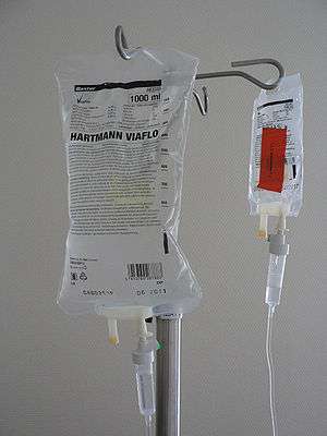
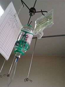
A standard IV infusion set consists of a pre-filled, sterile container (glass bottle, plastic bottle or plastic bag) of fluids with an attachment that allows the fluid to flow one drop at a time, making it easy to see the flow rate (and also reducing air bubbles); a long sterile tube with a clamp to regulate or stop the flow; a connector to attach to the access device; and Y-sets to allow "piggybacking" of another infusion set onto the same line, e.g., adding a dose of antibiotics to a continuous fluid drip.
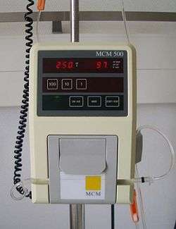
An infusion pump allows precise control over the flow rate and total amount delivered, but in cases where a change in the flow rate would not have serious consequences, or if pumps are not available, the drip is often left to flow simply by placing the bag above the level of the patient and using the clamp to regulate the rate; this is a gravity drip.
A rapid infuser can be used if the patient requires a high flow rate and the IV access device is of a large enough diameter to accommodate it. This is either an inflatable cuff placed around the fluid bag to force the fluid into the patient or a similar electrical device that may also heat the fluid being infused.
Intermittent infusion
Intermittent infusion is used when a patient requires medications only at certain times, and does not require additional fluid. It can use the same techniques as an intravenous drip (pump or gravity drip), but after the complete dose of medication has been given, the tubing is disconnected from the IV access device. Some medications are also given by IV push or bolus, meaning that a syringe is connected to the IV access device and the medication is injected directly (slowly, if it might irritate the vein or cause a too-rapid effect). Once a medicine has been injected into the fluid stream of the IV tubing there must be some means of ensuring that it gets from the tubing to the patient. Usually this is accomplished by allowing the fluid stream to flow normally and thereby carry the medicine into the bloodstream; however, a second fluid injection is sometimes used, a "flush", following the injection to push the medicine into the bloodstream more quickly.
History
Intravenous technology stems from studies on cholera treatment in 1831 by Dr Thomas Latta of Leith.[11]
Intravenous therapy was further developed in the 1930s by Hirschfeld, Hyman and Wanger[12][13] but was not widely available until the 1950s.[14]
Education
_(15836441352).jpg)
Intravenous therapy knowledge and skills among healthcare providers vary greatly. While initial exposure to I.V. therapy is usually through formal nursing education programs, I.V. starting skills only develop from a combination of theoretical instruction and on the job practice. However, employers usually expect potential employees to be proficient on this area of clinical practice prior to becoming hired. The gap between actual and expected knowledge and skills can be extremely wide, especially in I.V. therapy. Part of this gap comes from the lack of educational experiences and/or the inaccessibility to these offerings. While not widely offered, interactive multimedia I.V. education, such as hybrid courses combining online theory with hands-on practice, can provide a way to narrow that gap in a quick and cost-effective manner which will represent an improvement on patient safety and treatment outcomes.[15] Simulated intravenous solutions used for training purposes only have been manufactured; in at least one case, the routing of training solutions into a standard medical setting has led to patient adverse events.[16]
The Infusion Nurses Society offers comprehensive evidence-based educational resources for its members. The Infusion Nurses Certification Corporation offers the only nationally recognized and accredited certification for infusion nurse through the CRNI® credential.
See also
- Blood substitutes
- Blood transfusion
- Bolus (medicine)
- Dialysis
- Drip chamber
- Hypodermic needle
- Life support
- Oral rehydration therapy
- Saline flush
- Fluid warmer
References
- 1 2 3 An Update on Intravenous Fluids by Gregory S. Martin, MD, MSc
- ↑ "Systematic Review of Hypotonic Versus Isotonic Intravenous Fluids". 2013.
- ↑ Bentzer, P; Griesdale, DE; Boyd, J; MacLean, K; Sirounis, D; Ayas, NT (27 September 2016). "Will This Hemodynamically Unstable Patient Respond to a Bolus of Intravenous Fluids?". JAMA. 316 (12): 1298–309. PMID 27673307.
- 1 2 American College of Emergency Physicians, "Five Things Physicians and Patients Should Question", Choosing Wisely: an initiative of the ABIM Foundation, American College of Emergency Physicians, retrieved January 24, 2014, which cites
- Hartling, Lisa; Bellemare, Steven; Wiebe, Natasha; Russell, Kelly F; Klassen, Terry P; Craig, William Raine; Craig, William Raine (2006). "Oral versus intravenous rehydration for treating dehydration due to gastroenteritis in children". Cochrane Database Syst Rev: CD004390. doi:10.1002/14651858.CD004390.pub2. PMID 16856044.
- ↑ Griffith, RJ; Jordan, V; Herd, D; Reed, PW; Dalziel, SR (26 April 2016). "Vapocoolants (cold spray) for pain treatment during intravenous cannulation.". The Cochrane database of systematic reviews. 4: CD009484. doi:10.1002/14651858.CD009484.pub2. PMID 27113639.
- ↑ Schwamburger NT, Hancock RH, Chong CH, Hartup GR, Vandewalle KS (2012). "The rate of adverse events during IV conscious sedation". General dentistry. 60 (5): e341–4. PMID 23032244.
- ↑ Hadaway L (2007). "Infiltration and extravasation". American Journal of Nursing. 107 (8): 64–72. doi:10.1097/01.NAJ.0000282299.03441.c7. PMID 17667395.
- ↑ "Know The Difference: Infiltration vs. Extravasation" http://w3.rn.com/news/clinical_insights_details.aspx?Id=34318 Accessed 3/21/2014
- ↑ Jiha Ham (March 20, 2015). "A Life Upended After an IV Glucose Treatment Popular Among Asian Immigrants". The New York Times. Retrieved March 21, 2015.
Although many doctors warn Asian immigrants in New York that the effects of injecting glucose differ little from drinking sugary water, many Asians, especially of older generations, still use the intravenous solution. In their homelands, it is commonly prescribed by doctors as a method to cure colds, fevers and sometimes an upset stomach.
- ↑ CDC Morbidity and Mortality Weekly Report Aug 2002. "Guidelines for the Prevention of Intravascular Catheter-Related Infections". Retrieved 2008-03-13.
- ↑ MacGillivray, Neil (2009). "Dr Thomas Latta: the father of intravenous infusion therapy". Journal of Infection Prevention. 10 (Suppl. 1): 3–6. doi:10.1177/1757177409342141.
- ↑ Stanley, Autumn (1995). Mothers and daughters of invention: notes for a revised history of technology. Rutgers University Press. pp. 141–142. ISBN 978-0-8135-2197-8. Retrieved 2011-06-05.
Wanger and colleagues had in effect invented the modern I.V.-drip method of drug delivery [...]
- ↑ Hirschfeld, Samuel; Hyman, Harold Thomas; Wanger, Justine J. (February 1931). "Influence of velocity on the response to intravenous injections". Archives of Internal Medicine. 47 (2): 259–287. doi:10.1001/archinte.1931.00140200095007. Retrieved 2011-06-05.
- ↑ Laura Geggel (3 December 2012), A Royal Spotlight on a Rare Condition The New York Times
- ↑ "Intravenous Therapy Certification". Coral Gables, Florida, United States: National Healthcare Institute. Retrieved 2015-01-31.
- ↑ "FDA's investigation into patients being injected with simulated IV fluids continues" (Press release). United States Food and Drug Administration. 30 January 2015. FDA warns health care professionals not to inject patients with IV solutions from Wallcur, of San Diego.
Further reading
External links
| Wikimedia Commons has media related to Intravenous therapy. |
- IVTEAM.com
- IV-Therapy.net
- , Intravenous Therapy.
| Emergency medicine | |
|---|---|
| Equipment |
|
| Drugs | |
| Organisations | |
| Courses / Life support |
|
| Scoring systems | |
| |
| Oral |
| -solution.jpg)     | ||||||||||||||||||||||||||||||||
|---|---|---|---|---|---|---|---|---|---|---|---|---|---|---|---|---|---|---|---|---|---|---|---|---|---|---|---|---|---|---|---|---|---|---|
| Ophthalmic, otologic, nasal |
| |||||||||||||||||||||||||||||||||
| Urogenital | ||||||||||||||||||||||||||||||||||
| Rectal (enteral) | ||||||||||||||||||||||||||||||||||
| Dermal |
| |||||||||||||||||||||||||||||||||
| ||||||||||||||||||||||||||||||||||