Meconium peritonitis
| Meconium peritonitis | |
|---|---|
| Classification and external resources | |
| Specialty | pediatrics |
| ICD-10 | P78.0 |
| ICD-9-CM | 777.6 |
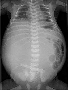
X-ray of a newborn with meconium pseudocyst resulting from bowel perforation. In this case the cause was atresia of the terminal ileum. There is a fine rim of calcification surrounding the big pseudocyst which shifts the other intestinal structures outwards.
Meconium peritonitis refers to rupture of the bowel prior to birth, resulting in fetal stool (meconium) escaping into the surrounding space (peritoneum) leading to inflammation (peritonitis). Despite the bowel rupture, many infants born after meconium peritonitis in utero have normal bowels and have no further issues.
Infants with cystic fibrosis are at increased risk for meconium peritonitis.
Diagnosis
Twenty percent of infants born with meconium peritonitis will have vomiting and dilated bowels on x-rays which necessitates surgery.
Meconium peritonitis is sometimes diagnosed on prenatal ultrasound[1] where it appears as calcifications[2] within the peritoneum.
History
Meconium peritonitis was first described in 1838 by Carl von Rokitansky.
References
- ↑ Tseng JJ, Chou MM, Ho ES (June 2003). "Meconium peritonitis in utero: prenatal sonographic findings and clinical implications". J Chin Med Assoc. 66 (6): 355–9. PMID 12889504.
- ↑ Dirkes, K; Crombleholme, TM; Craigo, SD; Latchaw, LA; Jacir, NN; Harris, BH; D'Alton, ME (July 1995). "The natural history of meconium peritonitis diagnosed in utero.". Journal of pediatric surgery. 30 (7): 979–82. doi:10.1016/0022-3468(95)90325-9. PMID 7472957.
External links
This article is issued from Wikipedia - version of the 5/27/2016. The text is available under the Creative Commons Attribution/Share Alike but additional terms may apply for the media files.