Placenta praevia
| Placenta previa | |
|---|---|
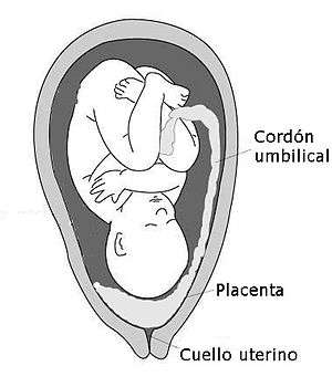 | |
| Diagram showing a placenta previa (Grade IV ) | |
| Classification and external resources | |
| Specialty | Obstetrics |
| ICD-10 | O44, P02.0 |
| ICD-9-CM | 641.0, 641.1 |
| MedlinePlus | 000900 |
| MeSH | D010923 |
Placenta previa is an obstetric complication in which the placenta is inserted partially or wholly in the lower uterine segment.[1] It is a leading cause of antepartum haemorrhage (vaginal bleeding). It affects approximately 0.4-0.5% of all labours.[2]
In the last trimester of pregnancy the isthmus of the uterus unfolds and forms the lower segment. In a typical pregnancy the placenta does not overlie. If the placenta does overlie the lower segment, as is the case with placenta previa, it may shear off and a small section may bleed.
Signs and symptoms
Women with placenta previa often present with painless, bright red vaginal bleeding. This commonly occurs around 32 weeks of gestation, but can be as early as late mid-trimester.[3] This bleeding often starts mildly and may increase as the area of placental separation increases. Previa should be suspected if there is bleeding after 24 weeks of gestation.
Women may also present as a case of failure of engagement of fetal head.[4]
Cause
Exact cause of placenta previa is unknown. It is hypothesized to be related to abnormal vascularisation of the endometrium caused by scarring or atrophy from previous trauma, surgery, or infection. These factors may reduce differential growth of lower segment, resulting in less upward shift in placental position as pregnancy advances.[5]
Classification
Traditionally, four grades of placenta previa were used,[1] however now it is more common to simply differentiate between 'major' and 'minor' cases.[6]
| Type | Description |
|---|---|
| Minor | Placenta is in lower uterine segment, but the lower edge does not cover the internal os |
| Major | Placenta is in lower uterine segment, and the lower edge covers the internal os |
Risk factors
| Risk factor | Odds ratio |
|---|---|
| Maternal age ≥ 40 (vs. < 20) | 9.1 |
| Illicit drugs | 2.8 |
| ≥ 1 previous Cesarean section | 2.7 |
| Parity ≥ 5 (vs. para 0) | 2.3 |
| Parity 2–4 (vs. para 0) | 1.9 |
| Prior abortion | 1.9 |
| Smoking | 1.6 |
| Congenital anomalies | 1.7 |
| Male fetus (vs. female) | 1.1 |
| Pregnancy-induced hypertension | 0.4 |
The following have been identified as risk factors for placenta previa:
- Previous placenta previa (recurrence rate 4–8%),[8] caesarean delivery,[9] myomectomy[4] or endometrium damage caused by D&C.[8]
- Women who are younger than 20 are at higher risk and women older than 35 are at increasing risk as they get older.
- Alcohol use during pregnancy was previous listed as a risk factor, but is discredited by this article.[10]
- Women who have had previous pregnancies, especially a large number of closely spaced pregnancies, are at higher risk due to uterine damage.[4]
- Smoking during pregnancy;[1] cocaine use during pregnancy[11][12]
- Women with a large placentae from twins or erythroblastosis are at higher risk.
- Race is a controversial risk factor, with some studies finding that people from Asia and Africa are at higher risk and others finding no difference.
- Placental pathology (Vellamentous insertion, succinturiate lobes, bipartite i.e. bilobed placenta etc.)[8]
- Baby is in an unusual position: breech (buttocks first) or transverse (lying horizontally across the womb).
Placenta previa is itself a risk factor of placenta accreta.
Diagnosis
History may reveal antepartum hemorrhage. Abdominal examination usually finds the uterus non-tender, soft and relaxed. Leopold's Maneuvers may find the fetus in an oblique or breech position or lying transverse as a result of the abnormal position of the placenta. Malpresentation is found in about 35% cases.[13] Vaginal examination is avoided in known cases of placenta previa.[1]
Confirmatory
Previa can be confirmed with an ultrasound.[14] Transvaginal ultrasound has superior accuracy as compared to transabdominal one, thus allowing measurement of distance between placenta and cervical os. This has rendered traditional classification of placenta previa obsolete.[15][16][17][18]
False positives may be due to following reasons:[19]
- Overfilled bladder compressing lower uterine segment
- Myometrial contraction simulating placental tissue in abnormally low location
- Early pregnancy low position, which in third trimester may be entirely normal due to differential growth of the uterus.
In such cases, repeat scanning is done after an interval of 15–30 minutes.
In parts of the world where ultrasound is unavailable, it is not uncommon to confirm the diagnosis with an examination in the surgical theatre. The proper timing of an examination in theatre is important. If the woman is not bleeding severely she can be managed non-operatively until the 36th week. By this time the baby's chance of survival is as good as at full term.
Management
An initial assessment to determine the status of the mother and fetus is required. Although mothers used to be treated in the hospital from the first bleeding episode until birth, it is now considered safe to treat placenta previa on an outpatient basis if the fetus is at less than 30 weeks of gestation, and neither the mother nor the fetus are in distress. Immediate delivery of the fetus may be indicated if the fetus is mature or if the fetus or mother are in distress. Blood volume replacement (to maintain blood pressure) and blood plasma replacement (to maintain fibrinogen levels) may be necessary.
The corticosteroids are indicated at 24–34 weeks gestation if the patient has bleeding, given the higher risk of premature birth. [Citation needed]
Mode of delivery
The mode of delivery is determined by clinical state of the mother, fetus and ultrasound findings. In minor degrees (traditional grade I and II), vaginal delivery is possible. RCOG recommends that the placenta should be at least 2 cm away from internal os for an attempted vaginal delivery.[20] When a vaginal delivery is attempted, consultant obstetrician and anesthetists are present in delivery suite. In cases of fetal distress and major degrees (traditional grade III and IV) a caesarean section is indicated. Caesarian section is contraindicated in cases of disseminated intravascular coagulation. An obstetrician may need to divide the anterior lying placenta. In such cases, blood loss is expected to be high and thus blood and blood products are always kept ready. In rare cases, hysterectomy may be required.[21] In the U.S., women with placenta previa who are covered by private insurance are 22% more likely to receive a caesarean section than women covered by Medicaid.[22]
Complications
Maternal
- Antepartum hemorrhage
- Malpresentation
- Abnormal placentation
- Postpartum hemorrhage
- Placenta previa increases the risk of puerperal sepsis and postpartum hemorrhage because the lower segment to which the placenta was attached contracts less well post-delivery.
Fetal
Epidemiology
Placenta previa occurs approximately one of every 250 births. One third of all antepartum hemorrhage occurs due to placenta previa. It has been suggested that incidence of placenta previa is increasing due to increased rate of Caesarian section.[23]
Perinatal mortality rate of placenta previa is 3-4 times higher than normal pregnancies.[24]
History
In places where a Caesarean section could not be performed due to the lack of a surgeon or equipment, infant could be delivered vaginally. There were two ways of doing this with a placenta previa:
- The baby's head can be brought down to the placental site (if necessary with Willet's forceps or a vulsellum) and a weight attached to its scalp
- A leg can be brought down and the baby's buttocks used to compress the placental site
The goal of this type of delivery is to save the mother, and both methods will often kill the baby. These methods were used for many years before Caesarean section and saved the lives of both mothers and babies with this condition. [Needs citations and expansion]
References
- 1 2 3 4 Arulkumaran, edited by Richard Warren, Sabaratnam (2009). Best practice in labour and delivery (1st ed., 3rd printing. ed.). Cambridge: Cambridge University Press. pp. 142–146. ISBN 978-0-521-72068-7.
- ↑ Faiz, AS; Ananth, CV (March 2003). "Etiology and risk factors for placenta previa: an overview and meta-analysis of observational studies.". The journal of maternal-fetal & neonatal medicine : the official journal of the European Association of Perinatal Medicine, the Federation of Asia and Oceania Perinatal Societies, the International Society of Perinatal Obstetricians. 13 (3): 175–90. doi:10.1080/jmf.13.3.175.190. PMID 12820840.
- ↑ Callander, Kevin P. Hanretty ; illustrated by Ian Ramsden, Robin (2004). Obstetrics illustrated (6th ed., Reprinted. ed.). Edinburgh [etc.]: Churchill Livingstone. p. 187. ISBN 0443072671.
- 1 2 3 Brinsden, Judith Collier, Murray Longmore, Mark (2006). Oxford handbook of clinical specialties (7th ed.). Oxford: Oxford University Press. p. 1970. ISBN 9780198530855.
- ↑ Dashe, JS; McIntire, DD; Ramus, RM; Santos-Ramos, R; Twickler, DM (May 2002). "Persistence of placenta previa according to gestational age at ultrasound detection.". Obstetrics and gynecology. 99 (5 Pt 1): 692–7. doi:10.1016/s0029-7844(02)01935-x. PMID 11978274.
- ↑ https://www.rcog.org.uk/en/guidelines-research-services/guidelines/gtg27/
- ↑ Jr, [edited by] E. Albert Reece, John C. Hobbins ; foreword by Norm F. Gant, (2006). Clinical obstetrics : the fetus and mother. (3 ed.). Malden, MA: Blackwell Pub. p. 1050. ISBN 978-1-4051-3216-9.
- 1 2 3 4 Kendrick, Chantal Simon, Hazel Everitt, Tony (2005). Oxford handbook of general practice (2nd ed.). Oxford: Oxford University Press. p. 793. ISBN 9780198565819.
- ↑ Weerasekera, D. S. (2000). "Placenta previa and scarred uterus - an obstetrician's dilemma". Journal of Obstetrics & Gynaecology. 20 (5): 484–5. doi:10.1080/014436100434659. PMID 15512632.
- ↑ Aliyu, MH; Lynch, O; Nana, PN; Alio, AP; Wilson, RE; Marty, PJ; Zoorob, R; Salihu, HM (July 2011). "Alcohol consumption during pregnancy and risk of placental abruption and placenta previa.". Maternal and child health journal. 15 (5): 670–6. doi:10.1007/s10995-010-0615-6. PMID 20437196.
- ↑ Handler, A; Kistin, N; Davis, F; Ferré, C (Apr 15, 1991). "Cocaine use during pregnancy: perinatal outcomes.". American Journal of Epidemiology. 133 (8): 818–25. PMID 2021149.
- ↑ Kistin, N; Handler, A; Davis, F; Ferre, C (July 1996). "Cocaine and cigarettes: a comparison of risks.". Paediatric and Perinatal Epidemiology. 10 (3): 269–78. doi:10.1111/j.1365-3016.1996.tb00050.x. PMID 8822770.
- ↑ Cotton, DB; Read, JA; Paul, RH; Quilligan, EJ (Jul 15, 1980). "The conservative aggressive management of placenta previa.". American Journal of Obstetrics and Gynecology. 137 (6): 687–95. doi:10.1016/s0002-9378(15)33242-7. PMID 7395932.
- ↑ Bhide, Amar; Thilaganathan, Basky (2004). "Recent advances in the management of placenta previa". Current Opinion in Obstetrics and Gynecology. 16 (6): 447–51. doi:10.1097/00001703-200412000-00002. PMID 15534438.
- ↑ Oppenheimer, LW; Farine, D; Ritchie, JW; Lewinsky, RM; Telford, J; Fairbanks, LA (October 1991). "What is a low-lying placenta?". American Journal of Obstetrics and Gynecology. 165 (4 Pt 1): 1036–8. doi:10.1016/0002-9378(91)90465-4. PMID 1951509.
- ↑ Neale, E. J.; Rogers, M. S. (1 July 1989). "Vaginal ultrasound for ruling out placenta previa. Case report". BJOG: an International Journal of Obstetrics and Gynaecology. 96 (7): 881–881. doi:10.1111/j.1471-0528.1989.tb03334.x.
- ↑ Smith, RS; Lauria, MR; Comstock, CH; Treadwell, MC; Kirk, JS; Lee, W; Bottoms, SF (January 1997). "Transvaginal ultrasonography for all placentas that appear to be low-lying or over the internal cervical os.". Ultrasound in Obstetrics & Gynecology. 9 (1): 22–4. doi:10.1046/j.1469-0705.1997.09010022.x. PMID 9060125.
- ↑ Farine, D; Fox, HE; Jakobson, S; Timor-Tritsch, IE (September 1988). "Vaginal ultrasound for diagnosis of placenta previa.". American Journal of Obstetrics and Gynecology. 159 (3): 566–9. doi:10.1016/s0002-9378(88)80009-7. PMID 3048096.
- ↑ Sutton, David (2003). Textbook of radiology and imaging (7th ed.). Edinburgh: Churchill Livingstone. p. 1064. ISBN 0443071098.
- ↑ "Placenta Previa, Placenta Previa Accreta and Vasa Previa: Diagnosis and Management". RCOG Guidelines - Green-top 27. Retrieved 15 January 2013.
- ↑ Kayem, G; Davy, C; Goffinet, F; Thomas, C; Clément, D; Cabrol, D (September 2004). "Conservative versus extirpative management in cases of placenta accreta.". Obstetrics and gynecology. 104 (3): 531–6. doi:10.1097/01.AOG.0000136086.78099.0f. PMID 15339764.
- ↑ Moore JE, Witt WP, Elixhauser A (April 2014). "Complicating Conditions Associate With Childbirth, by Delivery Method and Payer, 2011.". HCUP Statistical Brief #173. Rockville, MD: Agency for Healthcare Research and Quality.
- ↑ Miller, DA; Chollet, JA; Goodwin, TM (July 1997). "Clinical risk factors for placenta previa-placenta accreta.". American Journal of Obstetrics and Gynecology. 177 (1): 210–4. doi:10.1016/s0002-9378(97)70463-0. PMID 9240608.
- ↑ Crane, JM; van den Hof, MC; Dodds, L; Armson, BA; Liston, R (April 1999). "Neonatal outcomes with placenta previa". Obstetrics and gynecology. 93 (4): 541–4. doi:10.1016/s0029-7844(98)00480-3. PMID 10214830.