Ribonucleoprotein

Ribonucleoprotein (RNP) is a nucleoprotein that contains RNA, i.e. it is an association that combines a ribonucleic acid and an RNA-binding protein together. Such a combination can also be referred to as a protein-RNA complex. These complexes play an integral part in a number of important biological functions that include DNA replication, regulating gene expression[2] and regulating the metabolism of RNA.[3] A few examples of RNPs include the ribosome, the enzyme telomerase, vault ribonucleoproteins, RNase P, hnRNP and small nuclear RNPs (snRNPs), which have been implicated in pre-mRNA splicing (spliceosome) and are among the main components of the nucleolus.[4]
Currently, over 2000 RNPs can be found in the RCSB Protein Data Bank (PDB).[5] Furthermore, the Protein-RNA Interface Data Base (PRIDB) possesses a collection of information on RNA-protein interfaces based on data drawn from the PDB.[6] Some common features of protein-RNA interfaces were deduced based on known structures. For example, RNP in snRNPs have an RNA-binding motif in its RNA-binding protein. Aromatic amino acid residues in this motif result in stacking interactions with RNA. Lysine residues in the helical portion of RNA-binding proteins help to stabilize interactions with nucleic acids. This nucleic acid binding is strengthened by electrostatic attraction between the positive lysine side chains and the negative nucleic acid phosphate backbones. Additionally, it is possible to model RNPs computationally.[7] Although computational methods of deducing RNP structures are less accurate than experimental methods, they provide a rough model of the structure which allows for predictions of the identity of significant amino acids and nucleotide residues. Such information helps in understanding the overall function the RNP.

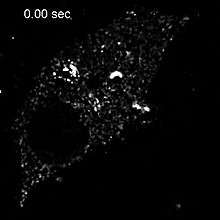
'RNP' can also refer to ribonucleoprotein particles. Ribonucleoprotein particles are distinct intracellular foci for post-transcriptional regulation. These particles play an important role in influenza A virus replication.[9] The influenza viral genome is composed of eight ribonucleoprotein particles formed by a complex of negative-sense RNA bound to a viral nucleoprotein. Each RNP carries with it an RNA polymerase complex. When the nucleoprotein binds to the viral RNA, it is able to expose the nucleotide bases which allow the viral polymerase to transcribe RNA. At this point, once the virus enters a host cell it will be prepared to begin the process of replication.
Anti-RNP antibodies
Anti-RNP antibodies are autoantibodies associated with mixed connective tissue disease and are also detected in nearly 40% of Lupus erythematosus patients. Two types of anti-RNP antibodies are closely related to Sjögren's syndrome: SS-A (Ro) and SS-B (La). Autoantibodies against snRNP are called Anti-Smith antibodies and are specific for SLE. The presence of a significant level of anti-U1-RNP also serves a possible indicator of MCTD when detected in conjunction with several other factors.[10]
List of RNPs
This is a (partial) list of ribonucleoprotein families:
- hnRNP
- Ribosome
- snRNP
- Signal recognition particle (a protein coated ribozyme)
- Telomerase
- Vault
References
- ↑ Muller, Mandy; Hutin, Stephanie; Marigold, Oliver; Li, Kathy H.; Burlingame, Al; Glaunsinger, Britt A. (2015-05-12). "A Ribonucleoprotein Complex Protects the Interleukin-6 mRNA from Degradation by Distinct Herpesviral Endonucleases". PLoS Pathogens. 11 (5). doi:10.1371/journal.ppat.1004899. ISSN 1553-7366. PMC 4428876
 . PMID 25965334.
 - ↑ Hogan, Daniel J; Riordan, Daniel P; Gerber, André P; Herschlag, Daniel; Brown, Patrick O (2016-11-07). "Diverse RNA-Binding Proteins Interact with Functionally Related Sets of RNAs, Suggesting an Extensive Regulatory System". PLoS Biology. 6 (10). doi:10.1371/journal.pbio.0060255. ISSN 1544-9173. PMC 2573929
 . PMID 18959479.
 - ↑ Lukong, Kiven E.; Chang, Kai-wei; Khandjian, Edouard W.; Richard, Stéphane (2008-08-01). "RNA-binding proteins in human genetic disease". Trends in genetics: TIG. 24 (8): 416–425. doi:10.1016/j.tig.2008.05.004. ISSN 0168-9525. PMID 18597886.
- ↑ "Ribonucleoprotein". www.uniprot.org. Retrieved 2016-11-07.
- ↑ Bank, RCSB Protein Data. "RCSB Protein Data Bank - RCSB PDB".
- ↑ Lewis, Benjamin A.; Walia, Rasna R.; Terribilini, Michael; Ferguson, Jeff; Zheng, Charles; Honavar, Vasant; Dobbs, Drena (2016-11-07). "PRIDB: a protein–RNA interface database". Nucleic Acids Research. 39 (Database issue): D277–D282. doi:10.1093/nar/gkq1108. ISSN 0305-1048. PMC 3013700
 . PMID 21071426.
 - ↑ Tuszynska, Irina; Matelska, Dorota; Magnus, Marcin; Chojnowski, Grzegorz; Kasprzak, Joanna M.; Kozlowski, Lukasz P.; Dunin-Horkawicz, Stanislaw; Bujnicki, Janusz M. (2014-02-01). "Computational modeling of protein-RNA complex structures". Methods (San Diego, Calif.). 65 (3): 310–319. doi:10.1016/j.ymeth.2013.09.014. ISSN 1095-9130. PMID 24083976.
- ↑ Momose, Fumitaka; Sekimoto, Tetsuya; Ohkura, Takashi; Jo, Shuichi; Kawaguchi, Atsushi; Nagata, Kyosuke; Morikawa, Yuko (2011-06-22). "Apical Transport of Influenza A Virus Ribonucleoprotein Requires Rab11-positive Recycling Endosome". PLoS ONE. 6 (6). doi:10.1371/journal.pone.0021123. ISSN 1932-6203. PMC 3120830
 . PMID 21731653.
 - ↑ Baudin, F; Bach, C; Cusack, S; Ruigrok, R W (1994-07-01). "Structure of influenza virus RNP. I. Influenza virus nucleoprotein melts secondary structure in panhandle RNA and exposes the bases to the solvent.". The EMBO Journal. 13 (13): 3158–3165. ISSN 0261-4189. PMC 395207
 . PMID 8039508.
 - ↑ "Mixed Connective Tissue Disease (MCTD) | Cleveland Clinic". my.clevelandclinic.org. Retrieved 2016-11-07.
External links
- Ribonucleoproteins at the US National Library of Medicine Medical Subject Headings (MeSH)
- PRIDB Protein-RNA Interface Database