Pulmonary vein
| Pulmonary Veins | |
|---|---|
.svg.png)
 | |
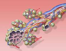
 Diagram of the alveoli with both cross-section and external view. | |
| Details | |
| Precursor | truncus arteriosus |
| Drains from | lungs |
| Drains to | left atrium |
| Artery | pulmonary artery |
| Identifiers | |
| Latin | venae pulmonales |
| MeSH | A07.231.908.713 |
| TA | A12.3.02.001 |
| FMA | 70827 |
The pulmonary veins are large blood vessels that receive oxygenated blood from the lungs and drain into the left atrium of the heart. There are four pulmonary veins, two from each lung. The pulmonary veins are among the few veins that carry oxygenated blood.
Structure
Two pulmonary veins emerge from each lung hilum, receiving blood from three or four bronchial veins apiece and draining into the left atrium. An inferior and superior vein drains each lung, so there are four veins in total.[1] The veins are fixed to the pericardium. The pulmonary veins travel alongside the pulmonary arteries[2]
At the root of the lung, the right superior pulmonary vein lies in front of and a little below the pulmonary artery; the inferior is situated at the lowest part of the lung hilum. Behind the pulmonary artery is the bronchus.[2]
The right pulmonary veins (contains oxygenated blood) pass behind the right atrium and superior vena cava; the left in front of the descending thoracic aorta.
Variation
Occasionally the three veins on the right side remain separate, and not infrequently the two left pulmonary veins end by a common opening into the left atrium. Therefore, the number of pulmonary veins opening into the left atrium can vary between three and five in the healthy population.
The two left pulmonary veins may be united as a single pulmonary vein in about 25% of people; the two right veins may be united in about 3%.[2]
Function
The pulmonary veins play an essential role in respiration, by receiving blood that has been oxygenated in the alveoli and returning it to the left atrium.
Clinical significance
As part of the pulmonary circulation they carry oxygenated blood back to the heart, as opposed to the veins of the systemic circulation which carry deoxygenated blood.
When the pulmonary veins drain into the systemic circulation in whole or in part, this is known as a total anomalous pulmonary venous circulation or partial anomalous pulmonary circulation, respectively.
Additional images
 Bronchial anatomy
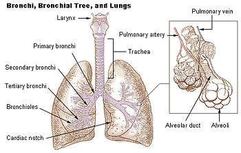 Bronchi, bronchial tree, and lungs
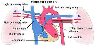 Pulmonary circuit
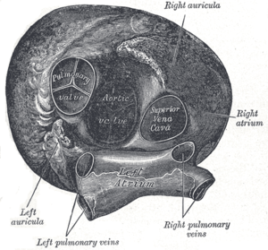 Heart seen from above.
 Transverse section of thorax, showing relations of pulmonary artery.
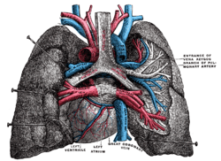 Pulmonary vessels, seen in a dorsal view of the heart and lungs.
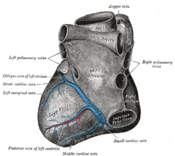 Base and diaphragmatic surface of heart.
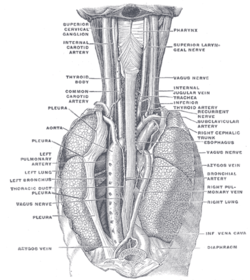 The position and relation of the esophagus in the cervical region and in the posterior mediastinum. Seen from behind.
 Left atrium
References
This article incorporates text in the public domain from the 20th edition of Gray's Anatomy (1918)
- ↑ Drake, Richard L.; Vogl, Wayne; Tibbitts, Adam W.M. Mitchell; illustrations by Richard; Richardson, Paul (2005). Gray's anatomy for students (Pbk. ed.). Philadelphia: Elsevier/Churchill Livingstone. ISBN 978-0-443-06612-2.
- 1 2 3 Skandalakis, editor in chief John E. (2004). "Chapter 7. Pericardium, Heart, and Great Vessels in the Thorax". Skandalakis' surgical anatomy : the embryologic and anatomic basis of modern surgery. Athens, Greece: PMP. pp. section titled 'Pulmonary veins'. ISBN 9603990744.
See also
External links
- Anatomy figure: 19:05-08 at Human Anatomy Online, SUNY Downstate Medical Center
- Illustration at infomat.net