Voxel-based morphometry
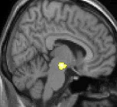
Voxel-based morphometry (VBM) is a neuroimaging analysis technique that allows investigation of focal differences in brain anatomy, using the statistical approach of statistical parametric mapping.
In traditional morphometry, volume of the whole brain or its subparts is measured by drawing regions of interest (ROIs) on images from brain scanning and calculating the volume enclosed. However, this is time consuming and can only provide measures of rather large areas. Smaller differences in volume may be overlooked. VBM registers every brain to a template, which gets rid of most of the large differences in brain anatomy among people. Then the brain images are smoothed so that each voxel represents the average of itself and its neighbors. Finally, the image volume is compared across brains at every voxel.
However, VBM can be sensitive to various artifacts, which include misalignment of brain structures, misclassification of tissue types, differences in folding patterns and in cortical thickness.[1] All these may confound the statistical analysis and either decrease the sensitivity to true volumetric effects, or increase the chance of false positives. For the cerebral cortex, it has been shown that volume differences identified with VBM may reflect mostly differences in surface area of the cortex, than in cortical thickness.[2][3]
History
One of the first VBM studies and one that came to attention in mainstream media was a study on the hippocampus brain structure of London taxicab drivers.[4] The VBM analysis showed the back part of the posterior hippocampus was on average larger in the taxi drivers compared to control subjects while the anterior hippocampus was smaller. London taxi drivers need good spatial navigational skills and scientists have usually associated hippocampus with this particular skill.
Another famous VBM paper was a study on the effect of age on gray and white matter and CSF of 465 normal adults.[5] The VBM analysis showed global gray matter was decreased linearly with age, especially for men, whereas global white matter did not decline with age.
A key description of the methodology of voxel-based morphometry is Voxel-Based Morphometry—The Methods[6] — one of the most cited articles in the journal NeuroImage.[7] The usual approach for statistical analysis is mass-univariate (analysis of each voxel separately), but pattern recognition may also be used, e.g., for classifying patients from healthy.[8]
VBM for brain asymmetry
Usually VBM is performed for examining differences across subjects, but it may also be used to examine neuroanatomical differences between hemispheres detecting brain asymmetry.[9][10] A technical procedure for such an investigation may use the following steps:[11]
- Construction of a study-specific brain image template with a balanced set of left and right handed and males and females.
- Construction of white and grey matter templates from segmentation.
- Construction of symmetric grey and white matter templates by averaging right and left cerebral hemispheres.
- Segmentation and extraction of brain image, e.g., removal of scalp tissue in the image.
- Spatial normalization to the symmetric templates
- Correction for volume change (applying a Jacobian determinant)
- Spatial smoothing.
- Actual statistical analysis by the general linear model, i.e., statistical parametric mapping.
See also
References
- ↑ John Ashburner (October 2001). "Computational anatomy with the SPM software". Magnetic Resonance Imaging. 27 (8): 1163–74. doi:10.1016/j.mri.2009.01.006. PMID 19249168.
- ↑ Natalie L. Voets; Morgan G. Hough; Gwenaëlle Douaud; Paul M. Matthews; Anthony James; Louise Winmill; Paula Webster; Stephen Smith (2008). "Evidence for abnormalities of cortical development in adolescent-onset schizophrenia". NeuroImage. 43 (4): 665–75. doi:10.1016/j.neuroimage.2008.08.013. PMID 18793730.
- ↑ Anderson M. Winkler; Peter Kochunov; John Blangero; Laura Almasy; Karl Zilles; Peter T. Fox; Ravindranath Duggirala; David C. Glahn (2010). "Cortical thickness or grey matter volume? The importance of selecting the phenotype for imaging genetics studies". NeuroImage. 53 (3): 1135–46. doi:10.1016/j.neuroimage.2009.12.028. PMC 2891595
 . PMID 20006715.
 - ↑ Eleanor A. Maguire, David G. Gadian, Ingrid S. Johnsrude, Catriona D. Good, John Ashburner, Richard S. J. Frackowiak, and Christopher D. Frith (2000). "Navigation-related structural change in the hippocampi of taxi drivers". Proceedings of the National Academy of Sciences. 97 (8): 4398–4403. Bibcode:2000PNAS...97.4398M. doi:10.1073/pnas.070039597. PMC 18253. PMID 10716738.
Commentary on the original article in the same issue:
- Alejandro Terrazas & Bruce L. McNaughton (2000). "Brain growth and the cognitive map". Proceedings of the National Academy of Sciences. 97 (9): 4414–4416. Bibcode:2000PNAS...97.4414T. doi:10.1073/pnas.97.9.4414. PMC 34310. PMID 10781031.
- BBC (2000-03-14). "Taxi drivers' brains 'grow' on the job". BBC News. Retrieved 2007-03-06.
- Alejandro Terrazas & Bruce L. McNaughton (2000). "Brain growth and the cognitive map". Proceedings of the National Academy of Sciences. 97 (9): 4414–4416. Bibcode:2000PNAS...97.4414T. doi:10.1073/pnas.97.9.4414. PMC 34310
- ↑ Catriona D. Good, Ingrid S. Johnsrude, John Ashburner, Richard N.A. Henson, Karl J. Friston and Richard S.J. Frackowiak (July 2001). "A Voxel-Based Morphometric Study of Ageing in 465 Normal Adult Human Brains" (PDF). NeuroImage. 14 (1): 21–36. doi:10.1006/nimg.2001.0786. PMID 11525331.
- ↑ John Ashburner and Karl J. Friston (June 2000). "Voxel-Based Morphometry—The Methods" (PDF). NeuroImage. 11 (6): 805–821. doi:10.1006/nimg.2000.0582. PMID 10860804.
- ↑ The number of citations is apparent from a search with Google Scholar (2007-12-07) .
- ↑ Yasuhiro Kawasaki, Michio Suzuki, Ferath Kherif, Tsutomu Takahashi, Shi-Yu Zhou, Kazue Nakamura, Mie Matsui, Tomiki Sumiyoshi, Hikaru Seto and Masayoshi Kurachi (January 2007). "Multivariate voxel-based morphometry successfully differentiates schizophrenia patients from healthy controls". NeuroImage. 34 (1): 235–242. doi:10.1016/j.neuroimage.2006.08.018. PMID 17045492.
- ↑ K.E. Watkins, T. Paus, J.P. Lerch, A. Zijdenbos, D.L. Collins, P. Neelin, J. Taylor, Keith J. Worsley and Alan C. Evans (September 2001). "Structural Asymmetries in the Human Brain: a Voxel-based Statistical Analysis of 142 MRI Scans". Cerebral Cortex. 11 (9): 868–877. doi:10.1093/cercor/11.9.868. PMID 11532891.
- ↑ E. Luders, C. Gaser, L. Jancke & G. Schlaug (June 2004). "A voxel-based approach to gray matter asymmetries". NeuroImage. 22 (2): 656–664. doi:10.1016/j.neuroimage.2004.01.032. PMID 15193594.
- ↑ Catriona D. Good, Ingrid Johnsrude, John Ashburner, Richard N. A. Henson, Karl J. Friston and Richard S. J. Frackowiak (September 2001). "Cerebral Asymmetry and the Effects of Sex and Handedness on Brain Structure: A Voxel-Based Morphometric Analysis of 465 Normal Adult Human Brains NeuroImage". NeuroImage. 14 (3): 685–700. doi:10.1006/nimg.2001.0857. PMID 11506541.
General technical references
- Tutorial: A Critical Analysis of Voxel Based Morphometry (VBM)
- Voxel-Based Morphometry Should Not Be Used with Imperfectly Registered Images
- Why Voxel-Based Morphometry Should Be Used
- Voxel Based Morphometry at the BIC
- Voxel-Based Morphometry at Wikibooks
- VBM tutorial by John Ashburner