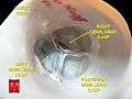
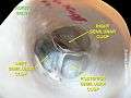
Aortic valve
| Aortic valve | |
|---|---|
.svg.png)
 Frontal view of the Aortic valve | |
 Aortic valve | |
| Details | |
| Identifiers | |
| Latin | valva aortae |
| TA | A12.1.04.012 |
| FMA | 7236 |
The aortic valve is a valve in the human heart between the left ventricle and the aorta. It is one of the two semilunar valves of the heart, the other being the pulmonary valve. The heart has four valves and the other two are the mitral and the tricuspid valves. The aortic valve normally has three cusps or leaflets, although in 1–2% of the population it is found to congenitally have two leaflets.[1]
Structure
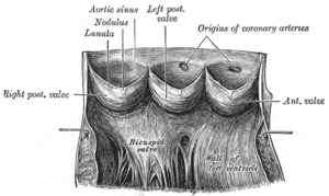
The aortic valve normally has three cusps – a left, right and posterior cusp.[2]
Function
When the left ventricle contracts (systole), pressure rises in the left ventricle. When the pressure in the left ventricle rises above the pressure in the aorta, the aortic valve opens, allowing blood to exit the left ventricle into the aorta. When ventricular systole ends, pressure in the left ventricle rapidly drops. When the pressure in the left ventricle decreases, the aortic pressure forces the aortic valve to close. The closure of the aortic valve contributes the A2 component of the second heart sound (S2).
Clinical significance

Narrowing of the aortic valve is called aortic stenosis, limiting the blood that can leave the valve and increasing the force the heart has to use to pump the blood through the valve. Aortic insufficiency, also called aortic regurgitation, is when the valve is unable to close properly. Blood consequently flows passively back to the heart in the wrong direction. These two conditions frequently co-exist. Common causes of aortic regurgitation include vasodilation of the aorta, previous rheumatic fever, infection such as infective endocarditis, degeneration of the aortic valve, and Marfan's syndrome. Aortic stenosis can also be caused by rheumatic fever and degenerative calcification.[3]
The most common congenital abnormality of the heart is the bicuspid aortic valve. In this condition, instead of three cusps, the aortic valve has two cusps. This condition is often undiagnosed until later in life when the person develops symptomatic aortic stenosis. Aortic stenosis occurs in this condition usually in patients in their 40s or 50s, an average of 10 years earlier than can occur in people with normal aortic valves.
Turner syndrome, a congenital condition that affects females, can often have a bicuspid aortic valve as one of its symptoms.
Aortic valve repair
Aortic valve repair or aortic valve reconstruction describes the reconstruction of both form and function of the native and dysfunctioning aortic valve. Most frequently it is applied for the treatment of aortic regurgitation. It can also become necessary for the treatment of aortic aneurysm, less frequently for congenital aortic stenosis.[4]
Aortic valve replacement
Aortic valve replacement is a surgical procedure in which a patient's aortic valve is replaced by a different valve. The aortic valve can be affected by a range of diseases and require aortic valve replacement. The valve can become either leaky (regurgitant or insufficient) or stuck partially shut (stenotic). Aortic valve replacement traditionally required open heart surgery. A new alternative is transcatheter aortic valve replacement (TAVR), which delivers a mechanical valve to the site of the diseased valve through a catheter.[5] There are two basic types of artificial heart valve, mechanical valves and tissue valves. Tissue heart valves are usually made from animal tissues, either animal heart valve tissue or animal pericardial tissue. The tissue is pretreated by removing antigens to prevent rejection and to prevent calcification.
There are alternatives to animal tissue valves. In some cases, a human aortic valve can be implanted. These are called homografts. Homograft valves are donated by patients and recovered after the patient expires. The durability of homograft valves is probably the same as for porcine tissue valves. Another procedure for aortic valve replacement is the Ross procedure (after Donald Ross) or pulmonary autograft. The Ross procedure involves going to surgery to have the aortic valve removed and replacing it with the patient's own pulmonary valve. A pulmonary homograft (a pulmonary valve taken from a cadaver) or a valvular prothesis is then used to replace the patient's own pulmonary valve.
The first minimally invasive aortic valve surgery took place at the Cleveland Clinic in 1996.
Another option for aortic valve replacement is transcatheter aortic valve replacement (TAVR). This procedure is for patients who are not candidates for surgery or who have high risk for surgery. This procedure can be done through the femoral artery, via direct aortic access, or via left ventricular apical access.
Additional images
-
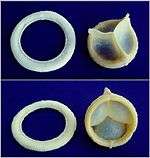
A replaceable model of Cardiac Biological Valve Prosthesis.
-
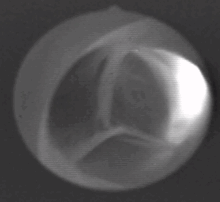
Aortic valves
-
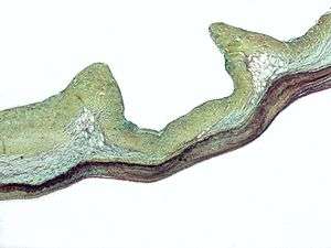
Aortic valve

References
- ↑ http://www.americanheart.org/presenter.jhtml?identifier=11068
- ↑ Anatomy photo:20:29-0104 at the SUNY Downstate Medical Center – "Heart: The Aortic Valve and Aortic Sinuses"
- ↑ Aortic Valve, Bicuspid at eMedicine
- ↑ Hans-Joachim Schäfers: Current treatment of aortic regurgitation. UNI-MED Science, Bremen, London, Boston 2013, ISBN 978-3-8374-1406-6.
- ↑ "What is TAVR?". American Heart Association. 2014. Retrieved 2015-08-15.
1. http://www.americanheart.org/presenter.jhtml?identifier=11068
External links
- Anatomy figure: 20:07-02 at Human Anatomy Online, SUNY Downstate Medical Center