Bird anatomy

Bird anatomy, or the physiological structure of birds' bodies, shows many unique adaptations, mostly aiding flight. Birds have a light skeletal system and light but powerful musculature which, along with circulatory and respiratory systems capable of very high metabolic rates and oxygen supply, permit the bird to fly. The development of a beak has led to evolution of a specially adapted digestive system. These anatomical specializations have earned birds their own class in the vertebrate phylum.
Skeletal system

1. skull
2. cervical vertebrae
3. furcula
4. coracoid
5. uncinate processes of ribs
6. keel
7. patella
8. tarsometatarsus
9. digits
10. tibia (tibiotarsus)
11. fibia (tibiotarsus)
12. femur
13. ischium (innominate)
14. pubis (innominate)
15. illium (innominate)
16. caudal vertebrae
17. pygostyle
18. synsacrum
19. scapula
20. lumbar vertebrae
21. humerus
22. ulna
23. radius
24. carpus (carpometacarpus)
25. metacarpus (carpometacarpus)
26. digits
27. alula
The bird skeleton is highly adapted for flight. It is extremely lightweight but strong enough to withstand the stresses of taking off, flying, and landing. One key adaptation is the fusing of bones into single ossifications, such as the pygostyle. Because of this, birds usually have a smaller number of bones than other terrestrial vertebrates. Birds also lack teeth or even a true jaw, instead having a beak, which is far more lightweight. The beaks of many baby birds have a projection called an egg tooth, which facilitates their exit from the amniotic egg, and that falls off once it has done its job.
Birds have many bones that are hollow (pneumatized) with criss-crossing struts or trusses for structural strength. The number of hollow bones varies among species, though large gliding and soaring birds tend to have the most. Respiratory air sacs often form air pockets within the semi-hollow bones of the bird's skeleton.[1]
The bones of diving birds are often less hollow than those of non-diving species. Loons[2] and puffins are without pneumatized bones entirely.[3] Flightless birds, such as ostriches and emus, demonstrate osseous pneumaticity, possessing pneumatized femurs[4] and, in the case of the emu, pneumatized cervical vertebrae.[5]
Birds also have more cervical (neck) vertebrae than many other animals; most have a highly flexible neck consisting of 13-25 vertebrae. Birds are the only vertebrates to have a fused collarbone (the furcula or wishbone) or a keeled sternum or breastbone. The keel of the sternum serves as an attachment site for the muscles used for flight or, similarly, for swimming, in penguins. Again, flightless birds, such as ostriches, which do not have highly developed pectoral muscles, lack a pronounced keel on the sternum. Swimming birds have a wide sternum, while walking birds have a long or high sternum and flying birds have a sternum width and height that are nearly equal.[6]
Birds have uncinate processes on the ribs. These are hooked extensions of bone which help to strengthen the rib cage by overlapping with the rib behind them. This feature is also found in the tuatara Sphenodon. They also have a greatly elongate tetradiate pelvis, similar to some reptiles. The hindlimb has an intra-tarsal joint found also in some reptiles. There is extensive fusion of the trunk vertebrae as well as fusion with the pectoral girdle. They have a diapsid skull, as in reptiles, with a pre-lachrymal fossa (present in some reptiles). The skull has a single occipital condyle.[7]
The skull consists of five major bones: the frontal (top of head), parietal (back of head), premaxillary and nasal (top beak), and the mandible (bottom beak). The skull of a normal bird usually weighs about 1% of the bird's total bodyweight. The eye occupies a considerable amount of the skull and is surrounded by a sclerotic eye-ring, a ring of tiny bones. This characteristic is also seen in reptiles.
The vertebral column consists of vertebrae, and is divided into three sections: cervical (11-25) (neck), synsacrum (fused vertebrae of the back, also fused to the hips (pelvis)), and pygostyle (tail).
The chest consists of the furcula (wishbone) and coracoid (collar bone), which, together with the scapula (see below), form the pectoral girdle. The side of the chest is formed by the ribs, which meet at the sternum (mid-line of the chest).
The shoulder consists of the scapula (shoulder blade), coracoid, and humerus (upper arm). The humerus joins the radius and ulna (forearm) to form the elbow. The carpus and metacarpus form the "wrist" and "hand" of the bird, and the digits are fused together. The bones in the wing are extremely light so that the bird can fly more easily.
The hips consist of the pelvis, which includes three major bones: the illium (top of the hip), ischium (sides of hip), and pubis (front of the hip). These are fused into one (the innominate bone). Innominate bones are evolutionary significant in that they allow birds to lay eggs. They meet at the acetabulum (hip socket) and articulate with the femur, which is the first bone of the hind limb.
The upper leg consists of the femur. At the knee joint, the femur connects to the tibiotarsus (shin) and fibula (side of lower leg). The tarsometatarsus forms the upper part of the foot, digits make up the toes. The leg bones of birds are the heaviest, contributing to a low center of gravity, which aids in flight. A bird's skeleton comprises only about 5% of its total body weight
Feet

Birds' feet are classified as anisodactyl, zygodactyl, heterodactyl, syndactyl or pamprodactyl.[8] Anisodactyl is the most common arrangement of digits in birds, with three toes forward and one back. This is common in songbirds and other perching birds, as well as hunting birds like eagles, hawks, and falcons.
Syndactyly, as it occurs in birds, is like anisodactyly, except that the third and fourth toes (the outer and middle forward-pointing toes), or three toes, are fused together, as in the belted kingfisher Ceryle alcyon. This is characteristic of Coraciiformes (kingfishers, bee-eaters, rollers, etc.).
The zygodactyly (from Greek ζυγον, a yoke) is an arrangement of digits in birds, with two toes facing forward (digits two and three) and two back (digits one and four). This arrangement is most common in arboreal species, particularly those that climb tree trunks or clamber through foliage. Zygodactyly occurs in the parrots, woodpeckers (including flickers), cuckoos (including roadrunners), and some owls. Zygodactyl tracks have been found dating to 120–110 Ma (early Cretaceous), 50 million years before the first identified zygodactyl fossils.[9]
Heterodactyly is like zygodactyly, except that digits three and four point forward and digits one and two point back. This is found only in trogons, while pamprodactyl is an arrangement in which all four toes may point forward, or birds may rotate the outer two toes backward. It is a characteristic of swifts (Apodidae).
Muscular system
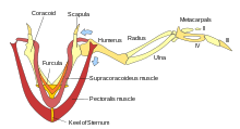
Most birds have approximately 175 different muscles, mainly controlling the wings, skin, and legs. The largest muscles in the bird are the pectorals, or the breast muscles, which control the wings and make up about 15 - 25% of a flighted bird's body weight. They provide the powerful wing stroke essential for flight. The muscle medial (underneath) to the pectorals is the supracoracoideus. It raises the wing between wingbeats. Both muscle groups attach to the keel of the sternum. This is remarkable, because other vertebrates have the muscles to raise the upper limbs generally attached to areas on the back of the spine. The supracoracoideus and the pectorals together make up about 25 – 35% of the bird's full body weight.
The skin muscles help a bird in its flight by adjusting the feathers, which are attached to the skin muscle and help the bird in its flight maneuvers.
There are only a few muscles in the trunk and the tail, but they are very strong and are essential for the bird. The pygostyle controls all the movement in the tail and controls the feathers in the tail. This gives the tail a larger surface area which helps keep the bird in the air.
Integumentary system

Scales
The scales of birds are composed of keratin, like beaks, claws, and spurs. They are found mainly on the toes and metatarsus, but may be found further up on the ankle in some birds. Most bird scales do not overlap significantly, except in the cases of kingfishers and woodpeckers. The scales and scutes of birds were originally thought to be homologous to those of reptiles and mammals;[10] however, more recent research suggests that scales in birds re-evolved after the evolution of feathers.[11][12]
Bird embryos begin development with smooth skin. On the feet, the corneum, or outermost layer, of this skin may keratinize, thicken and form scales. These scales can be organized into;
- Cancella – minute scales which are really just a thickening and hardening of the skin, crisscrossed with shallow grooves.
- Scutella – scales that are not quite as large as scutes, such as those found on the caudal, or hind part, of the chicken metatarsus.
- Scutes – the largest scales, usually on the anterior surface of the metatarsus and dorsal surface of the toes.
The rows of scutes on the anterior of the metatarsus can be called an "acrometatarsium" or "acrotarsium".
Reticula are located on the lateral and medial surfaces (sides) of the foot and were originally thought to be separate scales. However, histological and evolutionary developmental work in this area revealed that these structures lack beta-keratin (a hallmark of reptilian scales) and are entirely composed of alpha-keratin.[12] [13] This, along with their unique structure, has led to the suggestion that these are actually feather buds that were arrested early in development.[12]
Rhamphotheca and podotheca
The bills of many waders have Herbst corpuscles which help them find prey hidden under wet sand, by detecting minute pressure differences in the water.[14] All extant birds can move the parts of the upper jaw relative to the brain case. However this is more prominent in some birds and can be readily detected in parrots.[15]
The region between the eye and bill on the side of a bird's head is called the lore. This region is sometimes featherless, and the skin may be tinted, as in many species of the cormorant family.
The scaly covering present on the foot of the birds is called podotheca.
Beak
The beak, bill, or rostrum is an external anatomical structure of birds which is used for eating and for grooming, manipulating objects, killing prey, fighting, probing for food, courtship and feeding young. Although beaks vary significantly in size, shape and color, they share a similar underlying structure. Two bony projections—the upper and lower mandibles—covered with a thin keratinized layer of epidermis known as the rhamphotheca. In most species, two holes known as nares lead to the respiratory system.
Respiratory system

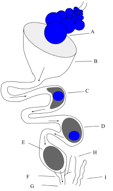
Due to their high metabolic rate required for flight, birds have a high oxygen demand. Their highly efficient respiratory system helps them meet that demand. Although birds have lungs they rely mostly on air sacs for ventilation. While bird lungs are smaller in comparison to mammals, the air sacs account for 15% of the total body volume, compared to 7% lung volume in mammals.[16]
The walls of these sacs do not have a good blood supply and so do not play a direct role in gas exchange. They act like a series of bellows[17] to move air unidirectionally through the respiratory system. Birds lack a diaphragm, so rather than the regular expansion and contraction of the respiratory organs as is seen in mammals, the air sacs allow the tract to maintain a fixed volume [18] with oxygenated air constantly flowing in a single direction through them.[19] The active phase of respiration in birds is exhalation, requiring muscular contraction.
Three distinct sets of organs perform respiration — the anterior air sacs (interclavicular, cervicals, and anterior thoracics), the lungs, and the posterior air sacs (posterior thoracics and abdominals). Typically there are nine air sacs within the system;[18] however, that number can range between seven and twelve, depending on the species of bird. Passeriformes possess seven air sacs, as the clavicular air sacs may interconnect or be fused with the cranial thoracic sacs. During inhalation, air initially enters the bird through the nares where it is heated, humidified, and filtered.[16] From there, the air enters the trachea and continues beyond the syrinx at which point the trachea branches into two bronchi, called the primary bronchi. The primary bronchi, or the mesobronchi, deliver the air to the posterior sacs at the caudal end of the bird. As the bird draws each breath, air is forced from the posterior air sacs, through the paleoparabronchi (commonly referred to as parabronchi) where gas exchange occurs, and then into the anterior sacs. Air from the anterior air sacs empties into the trachea and back out through the bird's mouth or nares during expiration.
The trachea is an area of dead space; air in the dead space is not fated to pass through the whole of the respiratory tract. In comparison to a mammalian respiratory tract, the dead space volume in a bird is 4.5 times greater than in mammals of the same size.[16] Birds with long necks, by association have long trachea and must compensate for higher dead space volumes.
Air passes through the lungs during both exhalation and inspiration, causing little to no mixing of new oxygen-rich air and stale carbon dioxide rich air as in mammalian lungs. Thus, the partial pressure of oxygen in a bird's respiratory tract is the same as the environment, and so birds have more efficient gas exchange than mammals do.
Avian lungs do not have alveoli as mammalian lungs do, but instead contain millions of tiny passages known as parabronchi, connected at both ends by the dorsobronchi and ventrobronchi. Air flows interiorly (caudal to cranial) through the parallel, honeycombed walls of the parabronchi into air vesicles, called atria, which project radially through the parabronchi. These atria give rise to air capillaries, where oxygen and carbon dioxide are exchanged with cross-flowing blood capillaries by diffusion.[20] All species of birds with the exception of the penguin, have neopulmonic parabronchi. These unorganized, unparalleled tubes project between the mesobronchus to the posterior sacs and into the posterior secondary bronchi. Unlike the paleoparabronchi, air traveling through the neopulmonic bronchi travels bidirectionally, compared to the unidirectional flow through the parabronchi. The neopulmonic parabronchi never make up more than 25% of the gas exchange surface.[16]
The syrinx is the sound-producing vocal organ of birds, located at the base of a bird's trachea. As with the mammalian larynx, sound is produced by the vibration of air flowing across the organ. The syrinx enables some species of birds to produce extremely complex vocalizations, even mimicking human speech. In some songbirds, the syrinx can produce more than one sound at a time.
Circulatory system
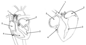
Birds have a four-chambered heart,[21] in common with humans, most mammals, and some reptiles (mainly the crocodilia). This adaptation allows for an efficient nutrient and oxygen transport throughout the body, providing birds with energy to fly and maintain high levels of activity. A ruby-throated hummingbird's heart beats up to 1200 times per minute (about 20 beats per second).[22]
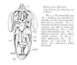
Digestive system
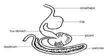
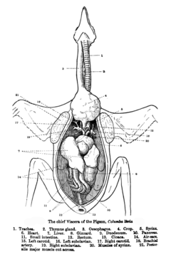
Many birds possess a muscular pouch along the esophagus called a crop. The crop functions to both soften food and regulate its flow through the system by storing it temporarily. The size and shape of the crop is quite variable among the birds. Members of the order Columbiformes, such as pigeons, produce a nutritious crop milk which is fed to their young by regurgitation. Birds possess a ventriculus, or gizzard, composed of four muscular bands that rotate and crush food by shifting the food from one area to the next within the gizzard. The gizzard of some species contains small pieces of grit or stone swallowed by the bird to aid in the grinding process of digestion, serving the function of mammalian or reptilian teeth. The use of gizzard stones is a similarity between birds and dinosaurs, which left gizzard stones called gastroliths as trace fossils.
Drinking behavior
There are four general ways in which birds drink: using gravity itself, sucking, use of the tongue, and deriving water entirely from food.
Most birds are unable to swallow by the "sucking" or "pumping" action of peristalsis in their esophagus (as humans do), and drink by repeatedly raising their heads after filling their mouths to allow the liquid to flow by gravity, a method usually described as "sipping" or "tipping up".[23] The notable exception is the Columbiformes; in fact, according to Konrad Lorenz in 1939:
one recognizes the order by the single behavioral characteristic, namely that in drinking the water is pumped up by peristalsis of the esophagus which occurs without exception within the order. The only other group, however, which shows the same behavior, the Pteroclidae, is placed near the doves just by this doubtlessly very old characteristic.[24]
Although this general rule still stands, since that time, observations have been made of a few exceptions in both directions.[23][25]
In addition, specialized nectar feeders like sunbirds (Nectariniidae) and hummingbirds (Trochilidae) drink by using protrusible grooved or trough-like tongues, and parrots (Psittacidae) lap up water.[23]
Many seabirds have glands near the eyes that allow them to drink seawater. Excess salt is eliminated from the nostrils. Many desert birds get the water that they need entirely from their food. The elimination of nitrogenous wastes as uric acid reduces the physiological demand for water.[26]
Reproductive and urogenital systems

Male birds have two testes which become hundreds of times larger during the breeding season to produce sperm.[27] The testes in male birds are generally asymmetric with most birds having a larger left testis.[28] Female birds in most families have only one functional ovary (the left one), connected to an oviduct — although two ovaries are present in the embryonic stage of each female bird. Some species of birds have two functional ovaries, and the order Apterygiformes always retain both ovaries.[29][30]
Most male birds have no phallus. In the males of species without a phallus, sperm is stored in the seminal glomera within the cloacal protuberance prior to copulation. During copulation, the female moves her tail to the side and the male either mounts the female from behind or in front (as in the stitchbird), or moves very close to her. The cloacae then touch, so that the sperm can enter the female's reproductive tract. This can happen very fast, sometimes in less than half a second.[31]
The sperm is stored in the female's sperm storage tubules for a period varying from a week to more than 100 days,[32] depending on the species. Then, eggs will be fertilized individually as they leave the ovaries, before the shell is calcified in the oviduct. After the egg is laid by the female, the embryo continues to develop in the egg outside the female body.

Many waterfowl and some other birds, such as the ostrich and turkey, possess a phallus. This appears to be the primitive condition among birds, most birds have lost the phallus.[33] The length is thought to be related to sperm competition in species that usually mate many times in a breeding season; sperm deposited closer to the ovaries is more likely to achieve fertilization.[34][35] The longer and more complicated phalli tend to occur in waterfowl whose females have unusual anatomical features of the vagina (such as dead end sacs and clockwise coils). These vaginal structures may be used to prevent penetration by the male phallus (which coils counter-clockwise). In these species, copulation is often violent and female co-operation is not required; the female ability to prevent fertilization may allow the female to choose the father for her offspring.[35][36][36][37][37][38] When not copulating, the phallus is hidden within the proctodeum compartment within the cloaca, just inside the vent.
After the eggs hatch, parents provide varying degrees of care in terms of food and protection. Precocial birds can care for themselves independently within minutes of hatching; altricial hatchlings are helpless, blind, and naked, and require extended parental care. The chicks of many ground-nesting birds such as partridges and waders are often able to run virtually immediately after hatching; such birds are referred to as nidifugous. The young of hole-nesters, on the other hand, are often totally incapable of unassisted survival. The process whereby a chick acquires feathers until it can fly is called "fledging".
Some birds, such as pigeons, geese, and red-crowned cranes, remain with their mates for life and may produce offspring on a regular basis.
Kidney
Avian kidneys function in almost the same way as the more extensively studied mammalian kidney, but with a few important adaptations; while much of the anatomy remains unchanged in design, some important modifications have occurred during their evolution. A bird has paired kidneys which are connected to the lower gastrointestinal tract through the ureters. Depending on the bird species, the cortex makes up around 71-80% of the kidney's mass, while the medulla is much smaller at about 5-15% of the mass. Blood vessels and other tubes make up the remaining mass. Unique to birds is the presence of two different types of nephrons (the functional unit of the kidney) both reptilian-like nephrons located in the cortex and mammalian-like nephrons located in the medulla. Reptilian nephrons are more abundant but lack the distinctive loops of Henle seen in mammals. The urine collected by the kidney is emptied into the cloaca through the ureters and then to the colon by reverse peristalsis.
Nervous system
Birds have acute eyesight—raptors have vision eight times sharper than humans—thanks to higher densities of photoreceptors in the retina (up to 1,000,000 per square mm in Buteos, compared to 200,000 for humans), a high number of neurons in the optic nerves, a second set of eye muscles not found in other animals, and, in some cases, an indented fovea which magnifies the central part of the visual field. Many species, including hummingbirds and albatrosses, have two foveas in each eye. Many birds can detect polarised light.
Birds have a large brain to body mass ratio. This is reflected in the advanced and complex bird intelligence.
Immune system
The immune system of birds resembles that of other animals. Birds have both innate and adaptive immume systems. Birds are susceptible to tumours, immune deficiency and autoimmune diseases.
See also
References
- ↑ Ritchison, Gary. "Ornithology (Bio 554/754):Bird Respiratory System". Eastern Kentucky University. Retrieved 2007-06-27.
- ↑ Gier, H. T. (1952). "The air sacs of the loon" (PDF). Auk. American Ornithologists' Union. 69: 40–49. doi:10.2307/4081291. Retrieved 2014-01-21.
- ↑ Fastovsky, David E.; Weishampel, David B. (2005). The Evolution and Extinction of the Dinosaurs (second ed.). Cambridge, New York, Melbourne,Madrid, Cape Town, Singapore, São Paulo: Cambridge University Press. ISBN 0-521-81172-4. Retrieved 2014-01-21.
- ↑ Bezuidenhout, A.J.; Groenewald, H.B.; Soley, J.T. (1999). "An anatomical study of the respiratory air sacs in ostriches" (PDF). Onderstepoort Journal of Veterinary Research. The Onderstepoort Veterinary Institute. 66: 317–325. Retrieved 2014-01-21.
- ↑ Wedel, Mathew J. (2003). "Vertebral pneumaticity, air sacs, and the physiology of sauropod dinosaurs" (PDF). Paleobiology. The Paleontological Society. 29 (2): 243–255. doi:10.1666/0094-8373(2003)029<0243:vpasat>2.0.co;2. Retrieved 2014-01-21.
- ↑ Duezler Ayhan; Ozgel Ozcan; Dursun Nejdet (2006). "Morphometric Analysis of the Sternum in Avian Species" (PDF). Turk. J. Vet. Anim. Sci. 30: 311–314.
- ↑ Wing, Leonard W. (1956) Natural History of Birds. The Ronald Press Company.
- ↑ Proctor, N. S. & Lynch, P. J. (1998) Manual of Ornithology: Avian Structure & Function. Yale University Press. ISBN 0300076193
- ↑ Lockley, M. G.; Li, R.; Harris, J. D.; Matsukawa, M.; Liu, M. (2007). "Earliest zygodactyl bird feet: Evidence from Early Cretaceous roadrunner-like tracks". Naturwissenschaften. 94 (8): 657–665. doi:10.1007/s00114-007-0239-x. PMID 17387416.
- ↑ Lucas, Alfred M. (1972). Avian Anatomy - integument. East Lansing, Michigan, USA: USDA Avian Anatomy Project, Michigan State University. pp. 67, 344, 394–601.
- ↑ Sawyer, R.H., Knapp, L.W. 2003. Avian Skin Development and the Evolutionary Origin of Feathers. J.Exp.Zool. (Mol.Dev.Evol) Vol.298B:57-72.
- 1 2 3 Dhouailly, D. 2009. A New Scenario for the Evolutionary Origin of Hair, Feather, and Avian Scales. J.Anat. Vol.214:587-606
- ↑ Stettenheim Peter R (2000). "The Integumentary Morphology of Modern Birds—An Overview". American Zoologist. 40 (4): 461–477. doi:10.1093/icb/40.4.461.
- ↑ Piersma, Theunis; Renee van Aelst; Karin Kurk; Herman Berkhoudt; Leo R. M. Maas (1998). "A New Pressure Sensory Mechanism for Prey Detection in Birds: The Use of Principles of Seabed Dynamics?". Proceedings: Biological Sciences. 265 (1404): 1377–1383. doi:10.1098/rspb.1998.0445.
- ↑ Zusi, R L (1984). "A Functional and Evolutionary Analysis of Rhynchokinesis in Birds.". Smithsonian Contributions to Zoology. 395. hdl:10088/5187.
- 1 2 3 4 Whittow, G. Causey (2000). Sturkie's Avian Physiology. San Diego, California: Academic Press. pp. 233–241. ISBN 978-0-12-747605-6.
- ↑ Calder, William A. (1996). Size, Function, and Life History. Mineola, New York: Courier Dove Publications. p. 91. ISBN 978-0-486-69191-6.
- 1 2 Krautwald-Junghanns, Maria-Elisabeth; et al. (2010). Diagnostic Imaging of Exotic Pets: Birds, Small Mammals, Reptiles. Germany: Manson Publishing. ISBN 978-3-89993-049-8.
- ↑ Ritchison, Gary. "Ornithology (Bio 554/754):Bird Respiratory System". Eastern Kentucky University. Retrieved 2007-06-27.
- ↑ "Bird lung". Archived from the original on March 11, 2007.
- ↑ Citation needed
- ↑ June Osborne (1998). The Ruby-Throated Hummingbird. University of Texas Press. p. 14. ISBN 0-292-76047-7.
- 1 2 3 Cade, Tom J. & Greenwald, Lewis I. (1966). "Drinking Behavior of Mousebirds in the Namib Desert, Southern Africa" (PDF). The Auk. 83 (1).
- ↑ K. Lorenz, Verhandl. Deutsch. Zool. Ges., 41 [Zool. Anz. Suppl. 12]: 69-102, 1939
- ↑ Cade, Tom J.; Willoughby, Ernest J. & Maclean, Gordon L. (1966). "Drinking Behavior of Sandgrouse in the Namib and Kalahari Deserts, Africa" (PDF). The Auk. 83 (1).
- ↑ Gordon L. Maclean (1996) The Ecophysiology of Desert Birds. Springer. ISBN 3-540-59269-5
- ↑ A study of the seasonal changes in avian testes Alexander Watson, J. Physiol. 1919;53;86-91, 'greenfinch (Carduelis chloris)', "In early summer (May and June) they are as big as a whole pea and in early winter (November) they are no bigger than a pin head"
- ↑ Lake, PE (1981). "Male genital organs". In King AS, McLelland J. Form and function in birds. 2. New York: Academic. pp. 1–61.
- ↑ Kinsky, FC (1971). "The consistent presence of paired ovaries in the Kiwi(Apteryx) with some discussion of this condition in other birds". Journal of Ornithology. 112 (3): 334–357. doi:10.1007/BF01640692.
- ↑ Fitzpatrick, FL (1934). "Unilateral and bilateral ovaries in raptorial birds" (PDF). Wilson Bulletin. 46 (1): 19–22.
- ↑ Lynch, Wayne; Lynch, photographs by Wayne (2007). Owls of the United States and Canada : a complete guide to their biology and behavior. Baltimore: Johns Hopkins University Press. p. 151. ISBN 0-8018-8687-2.
- ↑ Birkhead, TR; A. P. Moller (1993). "Sexual selection and the temporal separation of reproductive events: sperm storage data from reptiles, birds and mammals". Biological Journal of the Linnean Society. 50 (4): 295–311. doi:10.1111/j.1095-8312.1993.tb00933.x.
- ↑ Herrera, A. M; S. G. Shuster; C. L. Perriton; M. J. Cohn (2013). "Developmental Basis of Phallus Reduction during Bird Evolution". Current Biology. 23 (12): 1065–1074. doi:10.1016/j.cub.2013.04.062. PMID 23746636.
- ↑ McCracken, KG (2000). "The 20-cm Spiny Penis of the Argentine Lake Duck (Oxyura vittata)" (PDF). The Auk. 117 (3): 820–825. doi:10.1642/0004-8038(2000)117[0820:TCSPOT]2.0.CO;2.
- 1 2 Arnqvist, G.; I. Danielsson (1999). "Copulatory Behavior, Genital Morphology, and Male Fertilization Success in Water Striders". Evolution. 53: 147–156. doi:10.2307/2640927.
- 1 2 Eberhard, W (2010). "Evolution of genitalia: theories, evidence, and new directions". Genetica. 138: 5–18. doi:10.1007/s10709-009-9358-y.
- 1 2 Hosken, D.J.; P. Stockley (2004). "Sexual selection and genital evolution". Trends in Ecology & Evolution. 19: 87–93. doi:10.1016/j.tree.2003.11.012.
- ↑ Brennan, P. L. R.; R. O. Prum; K. G. McCracken; M. D. Sorenson; R. E. Wilson; T. R. Birkhead (2007). "Coevolution of Male and Female Genital Morphology in Waterfowl". PLOS ONE. 2: e418. doi:10.1371/journal.pone.0000418. PMC 1855079
 . PMID 17476339.
External links
Anatomy and morphology | ||
|---|---|---|
| Fields |
|  |
| Bacteria | ||
| Protists |
| |
| Plants |
| |
| Invertebrates | ||
| Mammals | ||
| Other vertebrates | ||
| Other topics | ||