Club foot
| Club foot | |
|---|---|
.jpg) | |
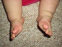
| Bilateral club foot | |
| Classification and external resources | |
| Specialty | Orthopedics |
| ICD-10 | M21.5, Q66.8 |
| ICD-9-CM | 736.71, 754.5-754.7 |
| OMIM | 119800 |
| DiseasesDB | 29395 |
| MedlinePlus | 001228 |
| eMedicine | radio/177 orthoped/598 |
| MeSH | D003025 |
Club foot or clubfoot, also called Congenital Talipes Equinovarus (CTEV), is a congenital deformity involving one foot or both.[1] The affected foot appears to have been rotated internally at the ankle. Without treatment, people with club feet often appear to walk on their ankles or on the sides of their feet. However, with treatment, the vast majority of patients recover completely during early childhood and are able to walk and participate in athletics just as well as patients born without CTEV.
It is a relatively common birth defect, occurring in about one in every 1,000 live births. Approximately half of people with clubfoot have it affect both feet, which is called bilateral club foot. In most cases it is an isolated disorder of the limbs. It occurs in males twice as frequently as in females.
A condition of the same name appears in some non-human animals, particularly horses, though in that particular case it is more akin to stepping en pointe than sideways.[2]
Genetics
Hippocrates around 400 B.C. was the first to offer a medical explanation, and to this day there are many hypotheses about how clubfoot develops, involving environmental factors, genetics, or a combination of both. No study has pinpointed the root cause, but most findings agree that "it is likely there is more than one different cause and at least in some cases the phenotype may occur as a result of a threshold effect of different factors acting together."[3]
Some researchers hypothesize, from the early development stages of humans, that clubfoot is formed by a malfunction during gestation. Amniocentesis is believed to increase the rates of this deformity because there is an increase in possible amniotic leakage during the procedure. Underdevelopment of the bones and muscles in the embryonic foot may be another underlying cause. Underdevelopment prevents the fetus's foot from rotating medially, leaving it in the club formation after birth. It was widely believed in the early 1900s that constriction of the foot by the uterus contributed to clubfoot.
Underdevelopment of the bones also affects the muscles and tissues of the foot. Abnormality in the connective tissue causes "the presence of increased fibrous tissue in muscles, fascia, ligaments and tendon sheaths".[3] Affected individuals have smaller than normal legs even after corrected. Further evidence shows that mutations in genes involved in muscle development are risk factors for clubfoot, specifically those encoding the muscle contractile complex (MYH3, TPM2, TNNT3, TNNI2, and MYH8). These can cause congenital contractures, including clubfoot, in distal arthrogryposis (DA) syndromes.[4] Clubfoot can also be present in patients with genetic conditions such as Loeys-Dietz syndrome.
Modern advances in genetic mapping techniques and the development of models of the disease in mice have improved understanding of developmental processes. Studies of genetic epidemiology have led to a more recent hypothesis on the etiology of clubfoot: its inheritance pattern is explained as a heterogenous disorder using a polygenic threshold model. The PITX1-TBX4 transcriptional pathway has become key to the study. PITX1 and TBX4 are uniquely expressed in the hind limb.[5] Further studies duplicating this pathway in models need to be conducted to hopefully find what genetic abnormalities cause clubfoot.
Diagnosis
Clubfoot is usually diagnosed immediately after birth simply by looking at the foot. It is then up to the doctor whether or not to x-ray the foot or feet to examine how the internal structures are positioned. In some cases, it may be possible to detect the disease prior to birth during the ultrasound. It may be more prominent if both feet are affected. The ability to possibly identify clubfoot before live birth can prove beneficial to the child as different treatments can be explored.[6]
Treatment
Once a child has been diagnosed with clubfoot, there are many different treatment approaches. Treatment should be given immediately after diagnosis to take full advantage of the flexibility in the baby’s bones and joints. This allows for improved manipulation to try to achieve a normal foot. The Ponseti method appears to result in better outcomes than the Kite method and similar outcomes to a traditional technique.[7]
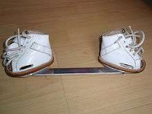
This involved manipulation by people specialized in the technique with serial casting and then providing braces to hold the feet in a plantigrade position. After serial casting, a foot abduction brace such as a Denis Browne bar with straight lace boots, ankle foot orthoses and/or custom foot orthoses (CFO) may be used. In North America, manipulation is followed by serial casting, most often by the Ponseti method. Foot manipulations usually begin within two weeks of birth.
French method
The French method, also known as the "functional method" or "physiotherapy method", is easiest to do with young bones. The child's foot is gradually stretched to achieve the right position, being held in place with tape after stretching. This method is not widely used , only in France, Texas Scottish Rite and a handful of other places. In the latest research publications issued in France, it is indicated that the French Functional method is more successful than the Ponseti method and that they will use this method countrywide.[8]
Extensive surgery of the soft tissue or bone is not usually necessary to treat clubfoot; however, a minimal procedure may be required:
- Tenotomy - clipping of the Achilles tendon – is needed in about 90% of cases in both the French and Ponseti method.[9][10]
In most cases extensive surgery is not needed to treat clubfoot. Extensive surgery may lead to scar tissue developing inside the child's foot. The scarring may result in functional, growth and aesthetic problems in the foot because the scarred tissue will interfere with the normal development of the appendage. A child who has extensive surgery may require on average two additional surgeries to correct the issues presented above.
Ponseti method

This treatment requires stretching. The foot is repositioned to the normal position then a cast (the "Ponseti cast") is placed on top of it. The baby’s foot is then continually repositioned and placed back into a cast once a week for several months. Towards the end of the process after being in a cast, the doctor will then surgically lengthen the heel cord (aka Achilles tendon). After the foot has been realigned, maintenance involves routine stretching. The child also has to wear special shoes or braces full-time for three months, then just nightly for three years. This method can be compared to wearing braces on your teeth. Parents have to follow the doctor’s orders for when to wear and not wear the brace to keep the foot corrected. Failure will occur if the brace is not worn and the foot will return to its odd shape.[6]
Treatment for clubfoot should begin almost immediately to have the best chance for a successful outcome without the need for surgery. Over the past 10 to 15 years, more and more success has been achieved in correcting clubfeet without the need for surgery. The clubfoot treatment method that is becoming the standard in the U.S. and worldwide is known as the Ponseti method.[11] Foot manipulations differ subtly from the Kite casting method which prevailed during the late 20th century. Although described by Dr. Ignacio Ponseti in the 1950s, it did not reach a wider audience until it was re-popularized around 2000 by Dr. John Herzenberg in the USA and in Europe and Africa by NHS surgeon Steve Mannion while working in Africa. Parents of children with clubfeet using the Internet also helped the Ponseti gain wider attention. The Ponseti method, if correctly done, is successful in >95% of cases[12] in correcting clubfeet using non- or minimal-surgical techniques. Typical clubfoot cases usually require five casts over four weeks. Atypical clubfeet and complex clubfeet may require a larger number of casts. Approximately 80% of infants require an Achilles tenotomy (microscopic incision in the tendon requiring only local anesthetic and no stitches) performed in a clinic toward the end of the serial casting.
After correction has been achieved, maintenance of correction may require the full-time (23 hours per day) use of a splint—also known as a foot abduction brace (FAB)—on both feet, regardless of whether the TEV is on one side or both, for several weeks after treatment. Part-time use of a brace (generally at night, usually 12 hours per day) is frequently prescribed for up to four years. Without the parents' participation, the clubfoot will almost certainly recur, because the muscles around the foot can pull it back into the abnormal position. Approximately 20% of infants successfully treated with the Ponseti casting method may require a surgical tendon transfer after two years of age. While this requires a general anesthetic, it is a relatively minor surgery that corrects a persistent muscle imbalance while avoiding disturbance to the joints of the foot. These treatments tend to be less effective in patients with Loeys-Dietz syndrome because of the underlying connective tissue disorder.
The common consensus among medical professionals regarding long-term outlook of club foot surgery is a full recovery and "normal" lifestyle. However, cases with no adverse impact long term are rare.
Botox is also being used as an alternative to surgery. Botox is the trade name for Botulinum Toxin type A, a chemical that acts on the nerves that control the muscle. It causes some paralysis (weakening) of the muscle by preventing muscle contractions (tightening). As part of the treatment for clubfoot, Botox is injected into the child’s calf muscle. In about one week, the Botox weakens the Achilles tendon. This allows the foot to be turned into a normal position over a period of 4–6 weeks, without surgery.
The weakness from a Botox injection usually lasts from 3–6 months. (Unlike surgery, it has no lasting effect). Most club feet can be corrected with just one Botox injection. It is possible to do another if it is needed. There is no scar or lasting damage.[13]
Surgery
In severe cases, surgery may be the only option to correct the foot after trying all other non-invasive methods for treatment. Surgery does not ensure full recovery, but most babies who underwent the surgery have maintained their normal feet. A surgeon will go in and lengthen the muscles and tendons to ease the foot into position. After surgery when the cast is removed, a brace is to be worn to prevent the foot from returning to the old position.[6]
On occasion, stretching, casting and bracing are not enough to correct a child's clubfoot. Surgery may be needed to adjust the tendons, ligaments and joints in the foot/ankle. Usually done at 9 to 12 months of age; surgery usually corrects all clubfoot deformities at the same time. After surgery, a cast holds the clubfoot still while it heals. It is still possible for the muscles in the child's foot to try to return to the clubfoot position, and special shoes or braces will likely be used for up to a year or more after surgery. Surgery will likely result in a stiffer foot than nonsurgical treatment, particularly over time.
Without any treatment, a child's clubfoot will result in severe functional disability, however with treatment, the child should have a nearly normal foot. He or she can run and play without pain and wear normal shoes. The corrected clubfoot will still not be perfect, however; a clubfoot usually stays 1 to 1½ sizes smaller and somewhat less mobile than a normal foot. The calf muscles in a leg with a clubfoot will also stay smaller. Many will require orthopedic shoes as adults, some will require a leg brace despite previous surgery.
Long-term studies of adults with post-club feet, especially those with substantial numbers of surgeries, may not fare as well in the long term, according to Dobbs, et al.,[14] A percentage of adults may require additional surgeries as they age, though there is some dispute as to the effectiveness of such surgeries, in light of the prevalence of scar tissue present from earlier surgeries.
In some cases the leg stops developing earlier than the healthy leg and a substantial length difference may occur. In some cases a leg lengthening will be necessary, most commonly by use of the Ilizarov method.
History
Treatment of clubfoot is evident as early as Egyptian paintings. In early days, the foot was manipulated with a Thomas wrench and casting which caused fracture of several bones in the foot.
Notable cases

Many notable people have been born with one or both feet in "clubbed" condition, including Roman emperor Claudius, statesman Prince Talleyrand, 19th-century American politician Thaddeus Stevens, comedian Damon Wayans, actor Gary Burghoff, and Eric The Midget from The Howard Stern Show, football players Steven Gerrard and Miguel Riffo, sledge hockey player Matt Lloyd, a Paralympian, mathematician Ben Greenberg, and filmmaker Jennifer Lynch.
While the British Romantic poet George Gordon, Lord Byron was wrongly said to suffer from clubfoot, he did suffer from a similar condition, tendon achillies, which caused him much humiliation.
Comedian, musician, and actor Dudley Moore was born with a club foot. This was mostly unknown to the public as he wore one shoe with a slightly bigger sole to compensate when walking.
NFL Cornerback Charles Woodson was born with severely clubbed feet and went on to win the Heisman Award at the University of Michigan, played in Super Bowl XXXVII with the Oakland Raiders and won Super Bowl XLV with the Green Bay Packers.
The figure-skater Kristi Yamaguchi was born with a clubfoot, and went on to win gold medals at both the 1992 Winter Olympics and World Championships. The soccer star Mia Hamm was born with the condition and won Gold twice with Team USA in the 1996 Olympics and in the 2004 Olympics. Baseball pitcher Larry Sherry, the 1959 World Series MVP, was born with club feet,[16] as was pitcher Jim Mecir, and both enjoyed long and successful careers. In fact, it was suggested in the book Moneyball that Mecir's club foot contributed to his success on the mound; it caused him to adopt a strange delivery that "put an especially violent spin" on his screwball, his specialty pitch. The San Francisco Giants held the record as the team with the all-time highest number of players with clubbed feet as of July 2010, and Freddy Sanchez, one of its infielders, cites his ability to overcome the defect as a reason for his success.[17] Tom Dempsey of the New Orleans Saints, born with a right club foot and no toes (this was his kicking foot), kicked an NFL record 63-yard (58 m) field goal. This kick became famous as the longest NFL field goal in history. Former NFL quarterback Troy Aikman beat being born with a clubfoot to enjoy a productive Hall of Fame career with 3 Super Bowl Rings in Super Bowl XXVII, Super Bowl XXVIII, and Super Bowl XXX.[18] Despite a club foot, Michael Houser, goaltender for the London Knights of the Ontario Hockey League, won the Red Tilson Trophy as the most outstanding player in the OHL in 2011-2012. He was signed by the National Hockey League's Florida Panthers in July, 2012.[19]
National Socialist and government official in Poland, Austria and Netherlands, Arthur Seyss-Inquart, lived his life walking with a clubfoot, and was still limping on it when he arrived at the gallows and was hanged as the last condemned prisoner at The Nuremberg Trials on October 16, 1946.[20] Nazi Propaganda Minister Joseph Goebbels had a deformity caused by a botched operation for the bacterial infection osteomyelitis, which some mistook for clubfoot.[21]
De Witt Clinton Fort, who served in the Confederate Army as a captain, was born with a clubfoot, and he was known during the American Civil War as Captain "Clubfoot" Fort, C.S.A.
Egyptian pharaoh Tutankhamun had a club foot and a cleft palate, and it is likely that he needed a cane to walk.[22]
In literature
- The main character, Philip Carey, in W. Somerset Maugham's novel Of Human Bondage, has a club foot, a central theme in the work.
- Hippolyte Tautain, the stable man at the Lion D'Or public house in Gustave Flaubert's novel Madame Bovary is unsuccessfully treated for clubfoot by Charles Bovary, leading to the eventual amputation of his leg.
- Charlie Wilcox, the main character in Sharon McKay's novel Charlie Wilcox had a club foot.
- In Yukio Mishima's seminal novel The Temple of the Golden Pavilion the character Kashiwagi has club feet which parallels the stutter of the main character, Mizoguchi.
- In David Eddings' Malloreon series, Senji the sorcerer has a club foot.
- In Caroline Lawrence's Roman Mysteries series, a character called Vulcan the blacksmith appears in the book "The Secrets of Vesuvius". He reveals that he gained the nickname because of his club foot.
- In Bernard Cornwell's The Warlord Chronicles Mordred, King of Dumnonia, has a club foot that is often used as a symbol for his ugliness and weakness as a ruler.
- In Daniel Keyes's Flowers for Algernon Gimpy, one of Charlie's co-workers at the bakery, has a club foot.
- In Perfume: The Story of a Murderer, the main character is born with a club foot and is described as having a limp throughout the novel.
- In Flannery O'Connor's short story "The Lame Shall Enter First", the character Johnson has a club foot, a major symbol within the story.
Other animals


Club feet occur in other animals, notably equines. The condition is characterized by a strongly upright pastern and a corresponding rotation of the coffin bone in the hoof. The condition often affects only one foot. Severity varies, with some animals usable for work or riding, and others unsound for life. Careful farrier work on the hooves can lessen the severity of many cases, and in certain circumstances surgery may be beneficial. The visible outward appearance of a club foot has different possible origins that include a genetic predisposition to the condition, a congenital defect formed while the animal is in the womb, or problems with diet and bone development during the early post-natal period. Certain horse breeds appear to be more predisposed to the condition than others, but research has yet to identify the genes involved.
A grading scale exists to assess the severity of club feet, which are caused by a deep digital flexor contraction syndrome. When the muscle fibers of the upper leg's deep digital flexor muscle contract excessively, this affects the tendon of the same name that inserts into this muscle group and attaches at the bottom of the coffin bone. A constant upward pull by the tendon on the coffin bone and other structure of the horse's hoof creates the condition. While many young foals are born with somewhat upright pasterns, the condition may resolve naturally or with minimal intervention if begun early. However, some cases are so severe that more drastic treatment may be required.[23]
References
- ↑ "CTEV: Deformities & Correction". LifeHugger. Retrieved 2009-12-26.
- ↑ Loving, Nancy S. "Managing the Club Foot". TheHorse.com. Retrieved 6 July 2016.
- 1 2 Miedzybrodzka, Z (January 2003). "Congenital talipes equinovarus (clubfoot): a disorder of the foot but not the hand.". Journal of Anatomy. 202 (1): 37–42. doi:10.1046/j.1469-7580.2003.00147.x. PMID 12587918.
- ↑ Weymouth, KS; Blanton, SH; Bamshad, MJ; Beck, AE; Alvarez, C; Richards, S; Gurnett, CA; Dobbs, MB; Barnes, D; Mitchell, LE; Hecht, JT (September 2011). "Variants in genes that encode muscle contractile proteins influence risk for isolated clubfoot.". American Journal of Medical Genetics Part A. 155A (9): 2170–9. doi:10.1002/ajmg.a.34167. PMID 21834041.
- ↑ Dobbs, MB; Gurnett, CA (January 2012). "Genetics of clubfoot.". Journal of pediatric orthopedics. Part B. 21 (1): 7–9. doi:10.1097/BPB.0b013e328349927c. PMID 21817922.
- 1 2 3 AskMayoExpert & et al. Can clubfoot be diagnosed in utero? Rochester, Minn.: Mayo Foundation for Medical Education and Research; 2012. http://www.mayoclinic.org/diseases-conditions/clubfoot/basics/definition/con-20027211
- ↑ Gray, K; Pacey, V; Gibbons, P; Little, D; Burns, J (Aug 12, 2014). "Interventions for congenital talipes equinovarus (clubfoot).". The Cochrane database of systematic reviews. 8: CD008602. doi:10.1002/14651858.CD008602.pub3. PMID 25117413.
- ↑ Islam, Muyedul; Ahmad, Akthar; Kabir, Rashedul; Bin Siddique, Kaoser; Arefin, Atia (May 2016). "Effectiveness of French Physiotherapy in Treating Congenital Clubfoot Deformity" (PDF). Orthopedics and Rheumatology. 3 (2). doi:10.19080/OROAJ.2016.02.555588. Retrieved 6 July 2016.
- ↑ "Tenotomy - procedure, pain, time, infection, operation, medication, risk, children". Surgery Encyclopedia. Retrieved 6 July 2016.
- ↑ S. Terry Canale; James H. Beaty (29 October 2012). Campbell's Operative Orthopaedics. Elsevier Health Sciences. pp. 997–. ISBN 0-323-08718-3.
- ↑ To Parents of Children Born with Clubfeet: Orthopaedics: UI Health Topics
- ↑ Morcuende JA, Dolan LA, Dietz FR, Ponseti IV (2004). "Radical reduction in the rate of extensive corrective surgery for clubfoot using the Ponseti method". Pediatrics. 113 (2): 376–80. doi:10.1542/peds.113.2.376. PMID 14754952.
- ↑ BC Women and Childrens Hospital
- ↑ Dobbs, Matthew B.; Nunley, R; Schoenecker, PL (May 2006). "Long-Term Follow-up of Patients with Clubfeet Treated with Extensive Soft-Tissue Release". The Journal of Bone & Joint Surgery (American). 88 (5): 986–96. doi:10.2106/JBJS.E.00114. PMID 16651573.
- ↑ Franck Fitoussi et Olivier Meslay, Un regard médicla sur le Piedbot, en collaboration avec l'hôpital Robert Debré sur le site cartelfr.louvre.fr
- ↑ The Big Book of Jewish Baseball: An ... - Google Books
- ↑ Kovacevic, Dejan (2006-08-18). "Freddy or not, here comes last leg of batting race". Pittsburgh Post-Gazette.
- ↑ Clubfoot doesn't stop rookie O.L. Simmons - New England Patriots Blog - ESPN Boston
- ↑ "Report: Top OHL goalie Houser headed to Florida - NHL.com - News". NHL.com. Retrieved 2013-07-05.
- ↑ http://digicoll.library.wisc.edu/cgi-bin/History/History-idx?type=turn&entity=History.Twilight.p0202&id=History.Twilight&isize=text&pview=hide
- ↑ Goebbels is commonly said to have had club foot (talipes equinovarus), a congenital condition. Historical writer and correspondent William L. Shirer spent the 1930s in Berlin as a journalist and was acquainted with Goebbels, wrote in his The Rise and Fall of the Third Reich (Simon and Schuster 1960) the Goebbels deformity arose from a childhood attack of osteomyelitis and a botched operation to correct it. Osteomyelitis, an infection within the bone marrow, can cause the destruction of one or more of the growing points in the long bones of the leg, a condition known as septic osteoblastic dysgenesis. This will result in a shortened leg.
- ↑ King Tut died from malaria, broken leg
- ↑ Redden, R.F. "Inside the Club Foot" The Horse, online edition, May 1, 2008. Accessed February 4, 2011