Bundle of His
| Bundle of His | |
|---|---|
 Isolated heart conduction system showing bundle of His | |
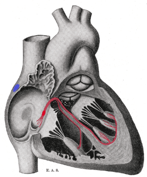
 Heart cut-away showing the bundle of His. Schematic representation of the atrioventricular bundle of His. The bundle, represented in red, originates near the orifice of the coronary sinus, undergoes slight enlargement to form the AV node. The AV node tapers down into the bundle of His, which passes into the ventricular septum and divides into two bundle branches, the left and right bundles. Sometimes the 'left and right bundles of His' are called Purkyně or Purkinje fibers. The ultimate distribution cannot be completely shown in this diagram. | |
| Details | |
| Identifiers | |
| Latin | fasciculus atrioventricularis |
| TA | A12.1.06.005 |
| FMA | 9484 |
The bundle of His or His bundle is a collection of heart muscle cells specialized for electrical conduction. As part of the electrical conduction system of the heart, it transmits the electrical impulses from the AV node (located between the atria and the ventricles) to the point of the apex of the fascicular branches via the bundle branches. The fascicular branches then lead to the Purkinje fibers, which provide electrical conduction to the ventricles, causing the cardiac muscle of the ventricles to contract at a paced interval.
Function
The bundle of His is an important part of the electrical conduction system of the heart, as it transmits impulses from the atrioventricular node, located at the inferior end of the interatrial septum, to the ventricles of the heart. The intrinsic rate of the bundle of His is 20 or less beats per minute.[1] The bundle of His branches into the left and the right bundle branches, which run along the interventricular septum. The left bundle branch further divides into the left anterior and the left posterior fascicles. These bundles and fascicles give rise to thin filaments known as Purkinje fibers. These fibers distribute the impulse to the ventricular muscle. The ventricular conduction system comprises the bundle branches and the Purkinje network. It takes about 0.03–0.04 seconds for the impulse to travel from the bundle of His to the ventricular muscle.
Clinical significance
Disorders affecting the cardiomyocytes that make up the electrical conduction system of the heart are called heart blocks. Heart blocks are separated into different categories based on the location of the cellular damage. Damage to any of the conducting cells in or below the bundle of His are collectively referred to as "infra-Hisian blocks". To be specific, blocks that occur in the right or left bundle branches are called "bundle branch blocks", and those that occur in either the left anterior or the left posterior fascicles are called "fascicular blocks", or "hemiblocks". The conditions in which both the right bundle branch and either the left anterior fascicle or the left posterior fascicle are blocked are collectively referred to as bifascicular blocks, and the condition in which the right bundle branch, the left anterior fascicle, and the left posterior fascicle are blocked is called trifascicular block. Infra-hisian blocks limit the heart's ability to coordinate the activities of the atria and ventricles, which usually results in a decrease in its efficiency in pumping blood.
Pacing
A 2000 study found that direct His bundle pacing is more effective in producing synchronized ventricular contraction—and therefore in improving cardiac function—than apical pacing.[2]
History
These specialized muscle fibers in the heart were named after the Swiss cardiologist Wilhelm His, Jr., who discovered them in 1893.[3][4]
See also
References
- ↑ Medical physiology; Boron
- ↑ Deshmukh P, Casavant DA, Romanyshyn M, Anderson K (February 2000). "Permanent, direct His-bundle pacing: a novel approach to cardiac pacing in patients with normal His-Purkinje activation". Circulation. 101 (8): 869–77. doi:10.1161/01.CIR.101.8.869. PMID 10694526.
- ↑ His' bundle at Who Named It?
- ↑ His, W., Jr. (1893). "Die Tätigkeit des embryonalen Herzens und deren Bedeutung für die Lehre von der Herzbewegung beim Erwachsenen". Arbeiten aus der medizinischen Klinik zu Leipzig. Jena, DE. 1: 14–50.
Further reading
- Scheinman MM, Saxon LA (February 2000). "Long-term His-bundle pacing and cardiac function". Circulation. 101 (8): 836–37. doi:10.1161/01.cir.101.8.836. PMID 10694517.
- Gillette PC, Swindle MM, Thompson RP, Case CL (April 1991). "Transvenous cryoablation of the bundle of His". Pacing and Clinical Electrophysiology. 14 (4 Pt 1): 504–10. PMID 1710054.
- Flowers NC, Hand RC, Orander PC, Miller CB, Walden MO, Horan LG (March 1974). "Surface recording of electrical activity from the region of the bundle of His". The American Journal of Cardiology. 33 (3): 384–89. doi:10.1016/0002-9149(74)90320-8. PMID 4591112.
- Kistin AD (May 1949). "Observations on the anatomy of the atrioventricular bundle (bundle of His) and the question of other muscular atrioventricular connections in normal human hearts". American Heart Journal. 37 (6): 847–67. doi:10.1016/s0002-8703(49)90937-0. PMID 18119894.
External links
- bundle of His at GPnotebook
- Atlas image: ht_rt_vent at the University of Michigan Health System - "Right atrioventricular bundle branch, anterior view"
- thoraxlesson4 at The Anatomy Lesson by Wesley Norman (Georgetown University) (thoraxheartinternalner)
- http://www.healthyheart.nhs.uk/heart_works/heart03.shtml