Ethmoid bone
| Ethmoid bone | |
|---|---|
 Cranial bones | |
 The seven bones which articulate to form the orbit. (Ethmoid is brown, between the red and the purple marked bones) | |
| Details | |
| Identifiers | |
| Latin | os ethmoidale |
| MeSH | A02.835.232.781.292 |
| TA | A02.1.07.001 |
| FMA | 52740 |
The ethmoid bone (/ˈɛθmɔɪd/;[1][2] from Greek ethmos, "sieve") is an unpaired bone in the skull that separates the nasal cavity from the brain. It is located at the roof of the nose, between the two orbits. The cubical bone is lightweight due to a spongy construction. The ethmoid bone is one of the bones that make up the orbit of the eye.
Structure
The ethmoid bone is an anterior cranial bone located between the eyes.[3] It contributes to the medial wall of the orbit, the nasal cavity, and the nasal septum.[4] The ethmoid has three parts: cribriform plate, ethmoidal labyrinth, and perpendicular plate. The cribriform plate forms the roof of the nasal cavity,the ethmoidal labyrinth consists of a large mass on either side of the perpendicular plate, and the perpendicular plate forms the superior two-thirds of the nasal septum.[5] Between the orbital plate and the nasal conchae are the ethmoidal sinuses or ethmoidal air cells, which are a variable number of small cavities in the lateral mass of the ethmoid.[6][7]
Articulations
The ethmoid articulates with thirteen bones:
- two bones of the neurocranium—the frontal, and the sphenoid (at the sphenoidal body and at the sphenoidal conchae).
- eleven bones of the viscerocranium—, two nasal bones, two maxillae, two lacrimals, two palatines, two inferior nasal conchae, and the vomer.
Development
The ethmoid is ossified in the cartilage of the nasal capsule by three centers: one for the perpendicular plate, and one for each labyrinth.
The labyrinths are first developed, ossific granules making their appearance in the region of the lamina papyracea between the fourth and fifth months of fetal life, and extending into the conchæ.
At birth, the bone consists of the two labyrinths, which are small and ill-developed. During the first year after birth, the perpendicular plate and crista galli begin to ossify from a single center, and are joined to the labyrinths about the beginning of the second year.
The cribriform plate is ossified partly from the perpendicular plate and partly from the labyrinths.
The development of the ethmoidal cells begins during fetal life.
Function
Role in magnetoception
Some birds and other migratory animals have deposits of biological magnetite in their ethmoid bones which allow them to sense the direction of the Earth's magnetic field. Humans have a similar magnetite deposit, but it is believed to be vestigial.[8]
Clinical significance
Fracture of the lamina papyracea, the lateral plate of the ethmoid labyrinth bone, permits communication between the nasal cavity and the ipsilateral orbit through the inferomedial orbital wall, resulting in orbital emphysema. Increased pressure within the nasal cavity, as seen during sneezing, for example, leads to temporary exophthalmos.
The porous fragile nature of the ethmoid bone makes it particularly susceptible to fractures. The ethmoid is usually fractured from an upward force to the nose. This could occur by hitting the dashboard in a car crash or landing on the ground after a fall. The ethmoid fracture can produce bone fragments that penetrate the cribriform plate. This trauma can lead to a leak of cerebrospinal fluid into the nasal cavity. These openings let opportunistic bacteria in the nasal cavity enter the sterile environment of the central nervous system (CNS). The CNS is usually protected by the blood-brain barrier, but holes in the cribriform plate let bacteria get through the barrier. The blood-brain barrier makes it extremely difficult to treat such infections, because only certain drugs can cross into the CNS.
An ethmoid fracture can also sever the olfactory nerve. This injury results in anosmia (loss of smell). A reduction in the ability to taste is also a side effect because it is based so heavily on smell. This injury is not fatal, but can be dangerous, as when a person fails to smell smoke, gas, or spoiled food.[9] In fact, people with anosmia were more than four times as likely to die in five years compared to those with a healthy sense of smell.[10]
Additional images
 Ethmoid bone from above.
 Perpendicular plate of ethmoid.
 Ethmoid bone (view from behind).
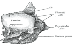 Ethmoid bone from the right side.
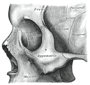 Ethmoid bone visible in the center, inside the orbit.
 Side view of the skull.
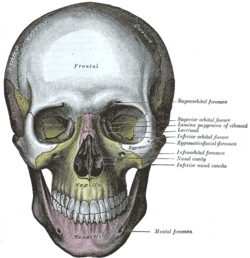 The skull from the front.
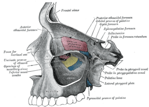 Medial wall of left orbit.
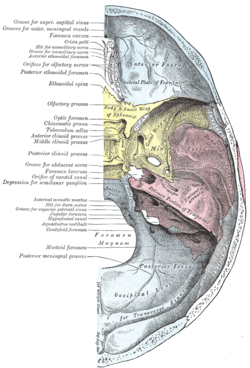 Base of the skull. Upper surface.
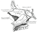 Medial wall of left nasal fossa.
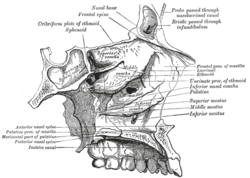 Roof, floor, and lateral wall of left nasal cavity.
 Lateral wall of nasal cavity, showing ethmoid bone in position.
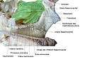 Ethmoid bone
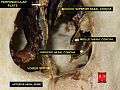 Perpendicular plate
See also
References
This article incorporates text in the public domain from the 20th edition of Gray's Anatomy (1918)
- ↑ OED 2nd edition, 1989 as /ˈεθmɔɪd/.
- ↑ Entry "ethmoid" in Merriam-Webster Online Dictionary.
- ↑ Saladin, Kenneth S. "Anatomy and Physiology: the Unity of Form and Function" McGraw Hill, 7th ed. New York: 2015
- ↑ Saladin, Kenneth S. "Anatomy and Physiology: the Unity of Form and Function" McGraw Hill, 7th ed. New York: 2015
- ↑ Saladin, Kenneth S. "Anatomy and Physiology: the Unity of Form and Function" McGraw Hill, 7th ed. New York: 2015
- ↑ Illustrated Anatomy of the Head and Neck, Fehrenbach and Herring, Elsevier, 2012, page 52
- ↑ Human Anatomy, Jacobs, Elsevier, 2008, page 210
- ↑ Nature 301, 78 - 80 (6 January 1983); doi:10.1038/301078a0
- ↑ Saladin, Kenneth S. "Anatomy and Physiology: the Unity of Form and Function" McGraw Hill, 7th ed. New York: 2015
- ↑ Pinto, Jayant, Kristen Wroblewski, David Kern, Phillip Schumm, and Martha McClintock. "Olfactory Dysfunction Predicts 5-Year Mortality in Older Adults." PLOS ONE:. 1 Oct. 2014. Web. 1 Dec. 2015.
Further reading
- Saladin, Kenneth S. "Anatomy and Physiology: the Unity of Form and Function" McGraw Hill, 5th ed. New York: 2010
- Banks, Peter; Brown, Andrew E. (2000). Fractures of the facial skeleton. Oxford: Wright. ISBN 0723610347. Retrieved 31 March 2012.
External links
| Wikimedia Commons has media related to Ethmoid bones. |
- http://www.theregister.com/2006/11/17/the_odd_body_nose_compass/
- Anatomy diagram: 34256.000-1 at Roche Lexicon - illustrated navigator, Elsevier