Nevus
| Nevus | |
|---|---|
|
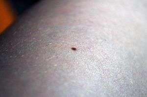
A mole on a leg | |
| Classification and external resources | |
| Specialty | dermatology |
| ICD-10 | I78.1 |
| ICD-9-CM | 448.1, 216.0-216.9 |
| MeSH | D009506 |
Nevus (plural: nevi) is a nonspecific medical term for a visible, circumscribed, chronic lesion of the skin or mucosa.[1] The term originates from nævus, which is Latin for "birthmark," however, a nevus can be either congenital (present at birth) or acquired. Common terms, including mole, birthmark, and beauty mark, are used to describe nevi, but these terms do not distinguish specific types of nevi.
Classification
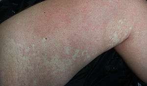
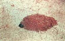
The term nevus is applied to a number of pigmentation disorders, both hypomelanotic and hypermelanotic,[2] as well as neoplasias and hyperplasias of melanocytes.[3]
Hypomelanotic

- Acquired
- Congenital
Hypermelanotic
.jpg)
- Acquired
- Acquired melanocytic nevus
- Junctional
- Intradermal
- Compound
- Atypical (dysplastic) nevus
- Becker's nevus
- Blue nevus (rarely congenital)
- Hori's nevus
- Nevus spilus
- Pigmented spindle cell nevus
- Spitz nevus
- Zosteriform lentiginous nevus
- Acquired melanocytic nevus
- Congenital

Diagnosis of nevi

Clinical diagnosis of a melanocytic nevus from other nevi can be made with the naked eye using the ABCD guideline, or using dermatoscopy. The main concern is distinguishing between a benign nevus, a dysplastic nevus, and a melanoma. Other skin tumors can resemble a melanocytic nevus clinically, such as a seborrheic keratosis, pigmented basal cell cancer, hemangiomas, and sebaceous hyperplasia. A skin biopsy is required when clinical diagnosis is inadequate or when malignancy is suspected.
Normal evolution or maturation of melanocytic nevi
All melanocytic nevi will change with time - both congenital and acquired nevi. The "normal" maturation is evident as elevation of the lesion from a flat macule to a raised papule. The color change occurs as the melanocytes clump and migrate from the surface of the skin (epidermis) down deep into the dermis. The color will change from even brown, to speckled brown, and then losing the color and becomes flesh colored or pink. During the evolution, uneven migration can make the nevi look like melanomas, and dermatoscopy can help in differentiation between the benign and malignant lesions.[4]
Etymology
A nevus may also be spelled naevus. The plural is nevi or naevi. The word is from nævus, Latin for "birthmark".
See also
References
- ↑ Happle, Rudolf (1995). "What is a nevus? A proposed definition of a common medical term.". Dermatology. 191(1): 1–5.
- ↑ "Chapter 75. Hypomelanoses and Hypermelanoses". Fitzpatrick's Dermatology in General Medicine. The McGraw Hill Companies, Inc. 2012. ISBN 978-0-07-166904-7.
- ↑ "Chapter 122. Benign Neoplasias and Hyperplasias of Melanocytes". Fitzpatrick's Dermatology in General Medicine. The McGraw-Hill Companies, Inc. 2012. ISBN 978-0-07-166904-7.
- ↑ Archived April 15, 2008, at the Wayback Machine.
External links
| Look up nevus in Wiktionary, the free dictionary. |
| Wikimedia Commons has media related to Nevus. |
- Atlas of Pathology Section of a melanocytic nevus
- eMedicine: Mole or Melanoma? Tell-Tale Signs in Benign Nevi and Malignant Melanoma: Slideshow
- Nevus Outreach, Inc.
- Pediatric plastic surgeon helps little girl with nevus NBC LA
