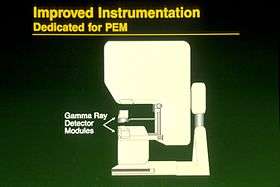
Positron emission mammography
| Positron emission mammography | |
|---|---|
| Diagnostics | |
 A positron emission mammography machine prototype | |
| ICD-10-PCS | CH3 |
Positron emission mammography (PEM) is a modality used to detect breast cancer.[1]
It is approved (by US FDA) for patients with a history of breast cancer.[2]
PEM uses a pair of gamma radiation detectors placed above and below the breast and mild breast compression to detect coincident gamma rays after administration of the radionuclide fluorine-18 fluorodeoxyglucose (Fludeoxyglucose (18F)), as used in whole-body PET studies for the detection of metastatic disease.[3]
References
- ↑ "Clinical imaging characteristics of the positron emission mammography camera: PEM Flex Solo II". J. Nucl. Med. 50 (10): 1666–75. October 2009. doi:10.2967/jnumed.109.064345. PMC 2873041
 . PMID 19759118.
 - ↑ Tartar, Marie; Comstock, Christopher E.; Kipper, Michael S. (2008). Breast cancer imaging: a multidisciplinary, multimodality approach. Elsevier Health Sciences. pp. 88–. ISBN 978-0-323-04677-0. Retrieved 18 July 2011.
- ↑ Glass and Shah (2013). "Clinical utility of positron emission mammography". Proc (Bayl Univ Med Cent). 26: 314–9. PMC 3684309
 . PMID 23814402.
![]() This article incorporates public domain material from the U.S. National Cancer Institute document "Dictionary of Cancer Terms".
This article incorporates public domain material from the U.S. National Cancer Institute document "Dictionary of Cancer Terms".
This article is issued from Wikipedia - version of the 10/12/2016. The text is available under the Creative Commons Attribution/Share Alike but additional terms may apply for the media files.