Myelography
| Myelography | |
|---|---|
| Intervention | |
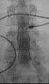
 Myelogram showing arachnoiditis in the lumbar spine. | |
| MeSH | D009192 |
| OPS-301 code | 3-130 |
Myelography is a type of radiographic examination that uses a contrast medium to detect pathology of the spinal cord, including the location of a spinal cord injury, cysts, and tumors. The procedure often involves injection of contrast medium into the cervical or lumbar spine, followed by several X-ray projections. A myelogram may help to find the cause of pain not found by an MRI or CT.
Myelography has been largely replaced by the use of CT and MRI scans.
Uses
A myelogram is sometimes used to better image the spinal cord in patients with lumbar spinal stenosis.[1]
Procedure
A CT is typically performed after radiographic contrast media (dye) has been placed with fluoroscopic guidance into a sac-like lining (the first- and hardest and outermost- layer of the spinal meninges, the spinal dura mater) surrounding the spinal cord and nerves. The material is typically water-based, which has largely replaced oil based fluids. A CT myelogram is most useful for patients who cannot undergo MRI (e.g., those with pacemakers or cochlear implants), or for those in whom MRI provides limited information (e.g., those with extensive metal in the spine).
The process usually involves lying face down on a table, with the lower extremities secured tightly with straps to the table. After the skin area has been numbed, the dye is injected into the thecal sac, then the table is slowly rotated in a circular motion, first down at the head end for approximately 4 to 6 minutes, then rotated up at the head end for the same duration. Several more minutes lying flat and the process is complete. This movement insures the contrast has sufficiently worked its way through the spinal cord, followed by X-rays, CT, or MRI scans.
If the fluid introduced in the spinal tap was oil based, the physician conducting the procedure will remove the fluid after the procedure is complete. When water-based fluid is used, it is typically not removed, as the fluid will eventually be absorbed into the body.
Post-procedure care centers around ensuring that infection (especially skin or subcutaneous infections, myelitis or meningitis or encephalitis, or sepsis) does not set in and that the "plug" at the site of the spinal tap does not become dislodged. Patients are usually instructed to avoid strenuous activity and heavy lifting, for example. Some patients are given instructions to keep their heads elevated at least 30 degrees for a specified number of hours. Complications from the surgery can cause a loss of cerebrospinal fluid (CSF), which could cause severe headaches. This can be corrected by returning to the medical facility and having them perform a blood patch. In this procedure a small amount of blood is taken from the arm and injected into the exact spinal tap location to stop the leaking of CSF.
 Myelography punction
 Conventional myelography in oblique projection. You can see the individual nerve root sheaths.
 Computed tomography after conventional myelography. The overlap-free representation often allows a more secure assessment. The high density of contrast material may be troublesome in case of insufficient mixing prior to CT.
References
- ↑ Katz, Jeffrey N.; Harris, Mitchel B. (February 21, 2008). "Lumbar Spinal Stenosis". New England Journal of Medicine. 358 (8): 818–825. doi:10.1056/NEJMcp0708097. PMID 18287604.
- Bontranger, Kenneth L. & Lampignano, John P. (2005). Radiographic Positioning and Related Anatomy, St. Louis: Elsevier Mosby. ISBN 0-323-02507-2.