Schlemm's canal
| Schlemm's canal | |
|---|---|
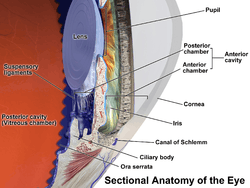 Anterior part of the human eye, with Canal of Schlemm at lower right. | |
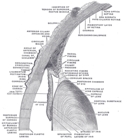 The upper half of a sagittal section through the front of the eyeball. (Canal of Schlemm labeled at center left.) | |
| Details | |
| Identifiers | |
| Latin | sinus venosus sclerae |
| TA | A12.3.06.109 |
| FMA | 51873 |
Schlemm's canal is a circular lymphatic-like vessel in the eye that collects aqueous humor from the anterior chamber and delivers it into the episcleral blood vessels via aqueous veins.[1][2][3][4] It is named after Friedrich Schlemm (1795–1858), a German anatomist.
The canal is essentially an endothelium-lined tube, resembling that of a lymphatic vessel. On the inside of the canal, nearest to the aqueous humor, it is covered by the trabecular meshwork; this region makes the greatest contribution to outflow resistance of the aqueous humor.
Conventionally, the canal has been considered a blood vessel, but recent studies have revealed that the molecular identity of Schlemm's canal is strikingly reminiscent to the one of lymphatic vasculature.[2][3][4]
Lymphatic-like identity
While conventional wisdom has considered Schlemm's canal (also known as the scleral venous sinus) as vein, the canal shares several structural and functional features reminiscent of the lymphatic vasculature. Notably, it is never filled with blood in physiological settings as it does not receive arterial blood circulation.[5]
Three recent independent pioneering studies by Aleksanteri Aspelund and Kari Alitalo from the University of Helsinki, Dae-Young Park and Gou Young Koh from KAIST and Krishnakumar Kizhatil and Simon W. M. John from the Howard Hughes Medical Institute, discovered that Schlemm's canal displays several features of lymphatic endothelium, including the expression of PROX1, VEGFR3, CCL21, FOXC2, but lacked the expression of LYVE1 and PDPN, indicating that Schlemm's canal is a lymphatic-like vessel.[2][3][4]
Developmental studies revealed that Schlemm's canal develops via a unique mechanism involving the transdifferentiation of venous endothelial cells in the eye into lymphatic-like endothelial cells.[2][3][4]
The developmental morphogenesis of the canal was sensitive to the inhibition of lymphangiogenic growth factors, and in adults, the administration of the lymphangiogenic growth factor VEGFC enlarged the Schlemm's canal, which was associated with a reduction in intraocular pressure.[2]
In the combined absence of angiopoietin 1 and angiopoietin 2, Schlemm's canal and episcleral lymphatic vasculature completely failed to develop, resulting in buphthalmos and glaucoma.[6]
Canaloplasty
Canaloplasty is a procedure designed to enhance and restore the eye’s natural drainage system to provide sustained reduction of intraocular pressure. Canaloplasty utilizes microcatheters in a simple and minimally invasive procedure. To perform a canaloplasty, a surgeon will create a tiny incision to gain access to Schlemm's canal. A microcatheter circumnavigates Schlemm's canal around the iris, enlarging the main drainage channel and its smaller collector channels through the injection of a sterile, gel-like material called viscoelastic. The catheter is then removed and a suture is placed within the canal and tightened. By opening Schlemm's canal, the pressure inside the eye is relieved. Long-term results are available, published in May 2009 in the Journal of Cataract and Refractive Surgery.[7][8][9][10]
Additional images
 Enlarged general view of the iridial angle. (Labeled with older label of 'sinus venosus scleræ' at center top.)
See also
References
- ↑ Cassin, B. and Solomon, S. Dictionary of Eye Terminology. Gainesville, Florida: Triad Publishing Company, 1990.
- 1 2 3 4 5 Aleksanteri Aspelund, Tuomas Tammela, Salli Antila, Harri Nurmi, Veli-Matti Leppänen, Georgia Zarkada, Lukas Stanczuk, Mathias Francois, Taija Mäkinen, Pipsa Saharinen, Ilkka Immonen, and Kari Alitalo. (2014). "The Schlemm's canal is a VEGF-C/VEGFR-3-responsive lymphatic-like vessel.". The Journal of Clinical Investigation. 124: 3975–86. doi:10.1172/JCI75395. PMID 25061878.
- 1 2 3 4 Dae-Young Park, Junyeop Lee, Intae Park, Dongwon Choi, Sunju Lee, Sukhyun Song, Yoonha Hwang, Ki Yong Hong, Yoshikazu Nakaoka, Taija Makinen, Pilhan Kim, Kari Alitalo, Young-Kwon Hong, and Gou Young Koh (2014). "Lymphatic regulator PROX1 determines Schlemm's canal integrity and identity.". The Journal of Clinical Investigation. 124: 3960–74. doi:10.1172/JCI75392. PMID 25061877.
- 1 2 3 4 Krishnakumar Kizhatil, Margaret Ryan, Jeffrey K. Marchant, Stephen Henrich, Simon W. M. John (2014). "Schlemm's canal is a unique vessel with a combination of blood vascular and lymphatic phenotypes that forms by a novel developmental process.". PLoS Biology. 12: e1001912. doi:10.1371/journal.pbio.1001912. PMC 4106723
 . PMID 25051267.
 - ↑ Ramos RF, Hoying JB, Witte MH, Daniel Stamer W. (2007). "Schlemm's canal endothelia, lymphatic, or blood vasculature?". J Glaucoma. 16: 391–405. doi:10.1097/IJG.0b013e3180654ac6. PMID 17571003.
- ↑ Thomson BR, Heinen S, Jeansson M, Ghosh AK, Fatima A, Sung HK, Onay T, Chen H, Yamaguchi S, Economides AN, Flenniken A, Gale NW, Hong YK, Fawzi A, Liu X, Kume T, Quaggin SE. (2014). "A lymphatic defect causes ocular hypertension and glaucoma in mice.". The Journal of Clinical Investigation. 124: 4320–4. doi:10.1172/JCI77162. PMID 25202984.
- ↑ https://mediamillinc.com/vjo.php?display=video&id=013
- ↑ http://www.jcrsjournal.org/article/S0886-3350(08)00004-7/abstract,
- ↑ http://www.jcrsjournal.org/article/S0886-3350(07)00697-9/abstract
- ↑ http://www.jcrsjournal.org/article/S0886-3350(09)00139-4/abstract
External links
- Histology image: 08005loa – Histology Learning System at Boston University
- Diagram
- Diagram
| Fibrous tunic (outer) |
| 
 | |||||||||
|---|---|---|---|---|---|---|---|---|---|---|---|
| Uvea/vascular tunic (middle) |
| ||||||||||
| Retina (inner) |
| ||||||||||
| Anatomical regions of the eye |
| ||||||||||
| Other | |||||||||||