Ranula
| Ranula | |
|---|---|
 | |
| Classification and external resources | |
| Specialty | otolaryngology |
| ICD-10 | K11.6 |
| ICD-9-CM | 527.6 |
| DiseasesDB | 31432 |
| MedlinePlus | 001639 |
| eMedicine | derm/648 |
| MeSH | D011900 |
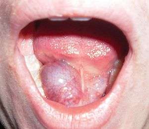
A ranula is a type of mucocele found on the floor of the mouth. Ranulas present as a swelling of connective tissue consisting of collected mucin from a ruptured salivary gland caused by local trauma. If small and asymptomatic further treatment may not be needed, otherwise minor oral surgery may be indicated.
Classification
A ranula is a type of mucocele, and therefore could be classified as a disorder of the salivary glands. Usually a ranula is confined to the floor of the mouth (termed a "simple ranula").[1] An unusual variant is the cervical ranula (also called a plunging or diving ranula), where the swelling is in the neck rather than the floor of the mouth.[2] The term ranula is also sometimes used to refer to other similar swellings of the floor of mouth such as true salivary duct cysts, dermoid cysts and cystic hygromas.[2] The Latin word rana means "frog" (ranula = "little frog"), and also now refers to a genus of frogs. The etymology of the term is usually explained by a resemblance with the underbelly,[2] or the bulging throat of a croaking frog.[3] Alternatively, ranula is described as being derived from the Greek word for "the swollen area below the mouth of a frog".[1]
Signs and symptoms

A ranula usually presents as a translucent blue, dome-shaped, fluctuant swelling in the tissues of the floor of the mouth. If the lesion is deeper, then there is a greater thickness of tissue separating from the oral cavity and the blue translucent appearance may not be a feature. A ranula can develop into a large lesion many centimeters in diameter, with resultant elevation of the tongue and possibly interfering with swallowing (dysphagia). The swelling is not fixed, may not show blanching and is non-painful unless it becomes secondarily infected. The usual location is usually is lateral to the midline, which may be used to help distinguish it from a midline dermoid cyst.[2] A cervical ranula presents as a swelling in the neck, with or without a swelling in the mouth. In common with other mucoceles, ranulas may rupture and then cause recurrent swelling. Ranulas may be asymptomatic, although they can fluctuate rapidly in size, shrinking and swelling, making them hard to detect.
Content of Ranula
Viscid and glairy of jelly like fluid.
Causes
Minor trauma to the floor of the mouth is thought to damage the delicate ducts that drain saliva from the sublingual gland into the oral cavity.[4] The lesion is a mucous extravasation cyst (mucocele) of the floor of mouth, although a ranula is often larger than other mucoceles (mainly because the overlying mucosa is thicker).[5] They can grow so large that they fill the mouth. The most usual source of the mucin spillage is the sublingual salivary gland, but ranulas may also arise from the submandibular duct or the minor salivary glands in the floor of the mouth. A cervical ranula occurs when the spilled mucin dissects its way through the mylohyoid muscle,[2] which separates the sublingual space from the submandibular space, and creates a swelling in the neck. It may occur following rupture of a simple ranula.[6] Rarely, ranulas may extend backwards into the parapharyngeal space.[6]
Diagnosis
The histologic appearance is similar to mucoceles from other locations. The spilled mucin causes a granulation tissue to form, which usually contains foamy histiocytes.[2] Ultrasound and magnetic resonance imaging may be useful to image the lesion.[6] A small squamous cell carcinoma obstructing Warton's duct may require clinical examination to be distinguished from a ranula.
Diagnostic criteria
- Mostly seen in young children and adolescents, both sexes are equally affected. Swelling in floor of mouth, which may be painful. Mostly unilateral, on one side of frenulum.
- Shape is spherical
- Size varies from 1 – 5 cm in diameter
- Color is pale blue with characteristics semi transparent appearance.
- Surface is smooth and mucous membrane is mobile over the swelling.
- Tenderness is absent
- Fluctuation test is positive
- Transillumination test is positive
- Cervical lymph nodes are not enlarged.
- May or may not have prolongation in the neck.
Treatment
Treatment of ranulas usually involves removal of the sublingual gland. Surgery may not be required if the ranula is small and asymptomatic.[4] Marsupialization may sometimes be used, where the intra-oral lesion is opened to the oral cavity with the aim of allowing the sublingual gland to re-establish connection with the oral cavity.
Complication
- Infection
- Repeated trauma
- Bursting and reformation
- Dysphagia in case of big Ranula
Epidemiology
The lesion is usually present in children.[4] Ranulas are the most common pathologic lesion associated with the sublingual glands.[5]
Other animals
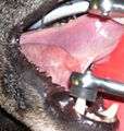
 Ranula in a dog
 excision of both mandibular and major sublingual glands in a dog
References
- Kahn, Michael A. Basic Oral and Maxillofacial Pathology. Volume 1. 2001.
- 1 2 Shaw, JHF. "Salivary Gland Surgery". unsupplied. Retrieved 8 February 2013.
- 1 2 3 4 5 6 Bouquot, Brad W. Neville , Douglas D. Damm, Carl M. Allen, Jerry E. (2002). Oral & maxillofacial pathology (2. ed.). Philadelphia: W.B. Saunders. pp. 391–392. ISBN 0721690033.
- ↑ Rao, PLNG. Problem Based Approach in Pediatric Surgery. New Delhi, India: Jaypee Brothers Publishers. p. 109. ISBN 8171795498.
- 1 2 3 Newlands, edited by Cyrus Kerawala, Carrie (2010). Oral and maxillofacial surgery. Oxford: Oxford University Press. p. 199. ISBN 9780199204830.
- 1 2 Hupp JR, Ellis E, Tucker MR (2008). Contemporary oral and maxillofacial surgery (5th ed.). St. Louis, Mo.: Mosby Elsevier. pp. 410–411. ISBN 9780323049030.
- 1 2 3 La'Porte, S. J.; Juttla, J. K.; Lingam, R. K. (14 September 2011). "Imaging the Floor of the Mouth and the Sublingual Space". Radiographics. 31 (5): 1215–1230. doi:10.1148/rg.315105062. PMID 21918039.
External links
| Wikimedia Commons has media related to Ranula. |