Acute coronary syndrome
| Acute coronary syndrome | |
|---|---|
|
Blockage of a coronary artery | |
| Classification and external resources | |
| Specialty | Cardiology |
| ICD-10 | I24.9 |
| eMedicine | emerg/31 |
| Patient UK | Acute coronary syndrome |
| MeSH | D054058 |
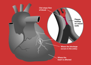
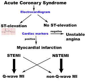
Acute coronary syndrome (ACS) is a syndrome (set of signs and symptoms) due to decreased blood flow in the coronary arteries such that part of the heart muscle is unable to function properly or dies.[1] The most common symptom is chest pain, often radiating to the left arm or angle of the jaw, pressure-like in character, and associated with nausea and sweating. Acute coronary syndrome is usually caused by one of three problems: ST elevation myocardial infarction (STEMI, 30%), non ST elevation myocardial infarction (NSTEMI, 25%), or unstable angina (38%).[2]
These types are named according to the appearance of the electrocardiogram (ECG/EKG) as non-ST segment elevation myocardial infarction and ST segment elevation myocardial infarction.[3] There can be some variation as to which forms of myocardial infarction (MI) are classified under acute coronary syndrome.[4]
ACS should be distinguished from stable angina, which develops during exertion and resolves at rest. In contrast with stable angina, unstable angina occurs suddenly, often at rest or with minimal exertion, or at lesser degrees of exertion than the individual's previous angina ("crescendo angina"). New onset angina is also considered unstable angina, since it suggests a new problem in a coronary artery.
Though ACS is usually associated with coronary thrombosis, it can also be associated with cocaine use.[5] Cardiac chest pain can also be precipitated by anemia, bradycardias (excessively slow heart rate) or tachycardias (excessively fast heart rate).
Signs and symptoms
The cardinal symptom of decreased blood flow to the heart is chest pain, experienced as tightness around the chest and radiating to the left arm and the left angle of the jaw. This may be associated with diaphoresis (sweating), nausea and vomiting, as well as shortness of breath. In many cases, the sensation is "atypical", with pain experienced in different ways or even being completely absent (which is more likely in female patients and those with diabetes). Some may report palpitations, anxiety or a sense of impending doom (angor animi) and a feeling of being acutely ill.
The description of the chest discomfort as a pressure has little utility in aiding a diagnosis as it is not specific for ACS.[6]
Diagnosis
Electrocardiogram
In the setting of acute chest pain, the electrocardiogram is the investigation that most reliably distinguishes between various causes.[8] The ECG should be done as early as practicable, including in the ambulance if possible.[9] If this indicates acute heart damage (elevation in the ST segment, new left bundle branch block), treatment for a heart attack in the form of angioplasty or thrombolysis is indicated immediately (see below). In the absence of such changes, it is not possible to immediately distinguish between unstable angina and NSTEMI.
Imaging and blood tests
As it is only one of the many potential causes of chest pain, the patient usually has a number of tests in the emergency department, such as a chest X-ray, blood tests (including myocardial markers such as troponin I or T, and H-FABP and/or a D-dimer if a pulmonary embolism is suspected), and telemetry (monitoring of the heart rhythm).
Prediction scores
The ACI-TIPI score can be used to aid diagnosis; using seven variables from the admission record, this score predicts crudely which patients are likely to have myocardial ischemia.[10] For example, according to a randomized controlled trial, males having chest pain with normal or non diagnostic ECG are at higher risk for having acute coronary syndrome than women.[11] In this study, the sensitivity was 65.2% and specificity was 44%. This particular study had an 8.4% prevalence of acute coronary syndrome, which means the positive predictive value of being a male with chest pain and having coronary syndrome is 9.6% and negative predictive value is 93.2% ( click here to adjust these results for patients at higher or lower risk of acute coronary syndrome).
In a second cohort study, exercise electrocardiography was similarly found to be a poor predictor of acute coronary syndrome at follow-up.[12] Of the patients who had a coronary event at 6 years of follow up, 47% had a negative ECG at the start of the study. With an average follow up of 2.21 years the receiver operating characteristic curves gave resting ECG a score of 0.72 and exercise ECG a score of 0.74.
There are not only prediction scores for diagnosis of ACS, but also prognosis. Most notably, the GRACE ACS Risk and Mortality score helps diagnose, and based upon that score predicts mortality rate of a given patient. It takes into account both clinical (blood pressure, heart rate, EKG findings) and medical history in its scoring system.[13]
Prevention
Acute coronary syndrome often reflects a degree of damage to the coronaries by atherosclerosis. Primary prevention of atherosclerosis is controlling the risk factors: healthy eating, exercise, treatment for hypertension and diabetes, avoiding smoking and controlling cholesterol levels; in patients with significant risk factors, aspirin has been shown to reduce the risk of cardiovascular events. Secondary prevention is discussed in myocardial infarction.
After a ban on smoking in all enclosed public places was introduced in Scotland in March 2006, there was a 17% reduction in hospital admissions for acute coronary syndrome. 67% of the decrease occurred in non-smokers.[14]
Treatment
People with presumed ACS are typically treated with aspirin, clopidogrel or ticagrelor, nitroglycerin, and if the chest discomfort persists morphine.[15] Other analgesics such as nitrous oxide are of unknown benefit.[15] Angiography is recommended in those who have either new ST elevation or a new left bundle branch block on their ECG.[1] Unless the person has low oxygen levels additional oxygen does not appear to be useful.[16]
STEMI
If the ECG confirms changes suggestive of myocardial infarction (ST elevations in specific leads, a new left bundle branch block or a true posterior MI pattern), thrombolytics may be administered or primary coronary angioplasty may be performed. In the former, medication is injected that stimulates fibrinolysis, destroying blood clots obstructing the coronary arteries. In the latter, a flexible catheter is passed via the femoral or radial arteries and advanced to the heart to identify blockages in the coronaries. When occlusions are found, they can be intervened upon mechanically with angioplasty and usually stent deployment if a lesion, termed the culprit lesion, is thought to be causing myocardial damage. Data suggest that rapid triage, transfer and treatment is essential.[17] The time frame for door-to-needle thrombolytic administration according to American College of Cardiology (ACC) guidelines should be within 30 minutes, whereas the door-to-balloon Percutaneous Coronary Intervention (PCI) time should be less than 90 minutes. It was found that thrombolysis is more likely to be delivered within the established ACC guidelines among patients with STEMI as compared to PCI according to a case control study.[18]
NSTEMI and NSTE-ACS
If the ECG does not show typical changes, the term "non-ST segment elevation ACS" is applied. The patient may still have suffered a "non-ST elevation MI" (NSTEMI). The accepted management of unstable angina and acute coronary syndrome is therefore empirical treatment with aspirin, a second platelet inhibitor such as clopidogrel, prasugrel or ticagrelor, and heparin (usually a low-molecular weight heparin such as enoxaparin), with intravenous glyceryl trinitrate and opioids if the pain persists.
A blood test is generally performed for cardiac troponins twelve hours after onset of the pain. If this is positive, coronary angiography is typically performed on an urgent basis, as this is highly predictive of a heart attack in the near-future. If the troponin is negative, a treadmill exercise test or a thallium scintigram may be requested.
If there is no evidence of ST segment elevation on the electrocardiogram, delaying urgent angioplasty until the next morning is not inferior to doing so immediately.[19] Using statins in the first 14 days after ACS reduces the risk of further ACS.[20]
In a cohort study comparing NSTEMI and STEMI, patients with NSTEMI had statistically similar mortality at one year after PCI as compared to patients with STEMI (3.4% vs 4.4%).[21] However, NSTEMI had significantly more "major cardiac events" (death, myocardial infarction, disabling stroke, or requiring revascularization) at one year (24.0% vs 16.6%).
Cocaine associated ACS should be managed in a manner similar to other patients with acute coronary syndrome except beta blockers should not be used and benzodiazepines should be administered early.[22]
Prognosis
TIMI score
The TIMI risk score can identify high risk patients[23] and has been independently validated.[24][25]
Biomarkers for diagnosis
The aim of diagnostic markers is to identify patients with ACS even when there is no evidence of heart muscle damage.
- Ischemia-Modified Albumin (IMA) – In cases of Ischemia – Albumin undergoes a conformational change and loses its ability to bind transitional metals (copper or cobalt). IMA can be used to assess the proportion of modified albumin in ischemia. Its use is limited to ruling out ischemia rather than a diagnostic test for the occurrence of ischemia.
- Myeloperoxidase (MPO) – The levels of circulating MPO, a leukocyte enzyme, elevate early after ACS and can be used as an early marker for the condition.
- Glycogen Phosphorylase Isoenzyme BB-(GPBB) is an early marker of cardiac ischemia and is one of three isoenzyme of Glycogen Phosphorylase.
- Troponin is a late cardiac marker of ACS
Biomarkers for risk determination
The aim of prognostic markers is to reflect different components of pathophysiology of ACS. For example:
- Natriuretic peptide – Both B-type natriuretic peptide (BNP) and N-terminal Pro BNP can be applied to predict the risk of death and heart failure following ACS.
- Monocyte chemo attractive protein (MCP)-1 – has been shown in a number of studies to identify patients with a higher risk of adverse outcomes after ACS.
Day of admission
Studies have shown that for ACS patients, weekend admission is associated with higher mortality and lower utilization of invasive cardiac procedures, and those who did undergo these interventions had higher rates of mortality and complications than their weekday counterparts. This data leads to the possible conclusion that access to diagnostic/interventional procedures may be contingent upon the day of admission, which may impact mortality.[26][27] This phenomenon is described as weekend effect.
References
- 1 2 Amsterdam, E. A.; Wenger, N. K.; Brindis, R. G.; Casey, D. E.; Ganiats, T. G.; Holmes, D. R.; Jaffe, A. S.; Jneid, H.; Kelly, R. F.; Kontos, M. C.; Levine, G. N.; Liebson, P. R.; Mukherjee, D.; Peterson, E. D.; Sabatine, M. S.; Smalling, R. W.; Zieman, S. J. (23 September 2014). "2014 AHA/ACC Guideline for the Management of Patients With Non-ST-Elevation Acute Coronary Syndromes: A Report of the American College of Cardiology/American Heart Association Task Force on Practice Guidelines". Circulation. 130 (25): e344–e426. doi:10.1161/CIR.0000000000000134. PMID 25249585.
- ↑ Torres M, Moayedi S (May 2007). "Evaluation of the acutely dyspneic elderly patient". Clin. Geriatr. Med. 23 (2): 307–25, vi. doi:10.1016/j.cger.2007.01.007. PMID 17462519.
- ↑ Grech ED, Ramsdale DR (June 2003). "Acute coronary syndrome: unstable angina and non-ST segment elevation myocardial infarction". BMJ. 326 (7401): 1259–61. doi:10.1136/bmj.326.7401.1259. PMC 1126130
 . PMID 12791748.
 - ↑ "Dorlands Medical Dictionary:acute coronary syndrome".
- ↑ Achar SA, Kundu S, Norcross WA (2005). "Diagnosis of acute coronary syndrome". Am Fam Physician. 72 (1): 119–26. PMID 16035692.
- ↑ Woo KM, Schneider JI (November 2009). "High-risk chief complaints I: chest pain--the big three". Emerg. Med. Clin. North Am. 27 (4): 685–712, x. doi:10.1016/j.emc.2009.07.007. PMID 19932401.
- ↑ Alpert JS, Thygesen K, Antman E, Bassand JP (2000). "Myocardial infarction redefined--a consensus document of The Joint European Society of Cardiology/American College of Cardiology Committee for the redefinition of myocardial infarction". J Am Coll Cardiol. 36 (3): 959–69. doi:10.1016/S0735-1097(00)00804-4. PMID 10987628.
- ↑ Chun AA, McGee SR (2004). "Bedside diagnosis of coronary artery disease: a systematic review". Am. J. Med. 117 (5): 334–43. doi:10.1016/j.amjmed.2004.03.021. PMID 15336583.
- ↑ Neumar, RW; Shuster, M; Callaway, CW; Gent, LM; Atkins, DL; Bhanji, F; Brooks, SC; de Caen, AR; Donnino, MW; Ferrer, JM; Kleinman, ME; Kronick, SL; Lavonas, EJ; Link, MS; Mancini, ME; Morrison, LJ; O'Connor, RE; Samson, RA; Schexnayder, SM; Singletary, EM; Sinz, EH; Travers, AH; Wyckoff, MH; Hazinski, MF (3 November 2015). "Part 1: Executive Summary: 2015 American Heart Association Guidelines Update for Cardiopulmonary Resuscitation and Emergency Cardiovascular Care.". Circulation. 132 (18 Suppl 2): S315–67. doi:10.1161/cir.0000000000000252. PMID 26472989.
- ↑ Selker HP, Griffith JL, D'Agostino RB (1991). "A tool for judging coronary care unit admission appropriateness, valid for both real-time and retrospective use. A time-insensitive predictive instrument (TIPI) for acute cardiac ischemia: a multicenter study". Medical care. 29 (7): 610–27. doi:10.1097/00005650-199107000-00002. PMID 2072767.
- ↑ Goodacre, S; Pett, P; Arnold, J; Chawla, A; Hollingsworth, J; Roe, D; Crowder, S; Mann, C; Pitcher, D; Brett, C (2009). "Clinical diagnosis of acute coronary syndrome in patients with chest pain and a normal or non-diagnostic electrocardiogram.". Emergency medicine journal : EMJ. 26 (12): 866–70. doi:10.1136/emj.2008.064428. PMID 19934131.
- ↑ Sekhri, N; Feder, GS; Junghans, C; Eldridge, S; Umaipalan, A; Madhu, R; Hemingway, H; Timmis, AD (2008). "Incremental prognostic value of the exercise electrocardiogram in the initial assessment of patients with suspected angina: cohort study.". BMJ(Clinical research ed.). 337: a2240. doi:10.1136/bmj.a2240. PMC 2583389. PMID 19008264.
- ↑ Fox KA, Dabbous OH, Goldberg RJ, Pieper KS, Eagle KA, Van de Werf F, Avezum A, Goodman SG, Flather MD, Anderson FA Jr, Granger CB (2006). "Prediction of risk of death and myocardial infarction in the six months after presentation with acute coronary syndrome: prospective multinational observational study (GRACE).". BMJ. 333 (7578): 1091. doi:10.1136/bmj.38985.646481.55. PMC 1661748. PMID 17032691.
- ↑ Pell JP, Haw S, Cobbe S, et al. (2008). "Smoke-free Legislation and Hospitalizations for Acute Coronary Syndrome". New England Journal of Medicine. 359 (5): 482–91. doi:10.1056/NEJMsa0706740. PMID 18669427.
- 1 2 O'Connor RE, Brady W, Brooks SC, et al. (November 2010). "Part 10: acute coronary syndromes: 2010 American Heart Association Guidelines for Cardiopulmonary Resuscitation and Emergency Cardiovascular Care". Circulation. 122 (18 Suppl 3): S787–817. doi:10.1161/CIRCULATIONAHA.110.971028. PMID 20956226.
- ↑ Neumar, RW; Shuster, M; Callaway, CW; Gent, LM; Atkins, DL; Bhanji, F; Brooks, SC; de Caen, AR; Donnino, MW; Ferrer, JM; Kleinman, ME; Kronick, SL; Lavonas, EJ; Link, MS; Mancini, ME; Morrison, LJ; O'Connor, RE; Samson, RA; Schexnayder, SM; Singletary, EM; Sinz, EH; Travers, AH; Wyckoff, MH; Hazinski, MF (3 November 2015). "Part 1: Executive Summary: 2015 American Heart Association Guidelines Update for Cardiopulmonary Resuscitation and Emergency Cardiovascular Care.". Circulation. 132 (18 Suppl 2): S315–67. doi:10.1161/cir.0000000000000252. PMID 26472989.
- ↑ Blankenship JC, Skelding KA (2008). "Rapid Triage, Transfer, and Treatment with Percutaneous Coronary Intervention for Patients with ST-Segment Elevation Myocardial Infarction". Acute Coronary Syndromes. 9 (2): 59–65.
- ↑ Janda, SP; Tan, N (2009). "Thrombolysis versus primary percutaneous coronary intervention for ST elevation myocardial infarctions at Chilliwack General Hospital.". The Canadian journal of cardiology. 25 (11): e382–4. doi:10.1016/S0828-282X(09)70165-5. PMC 2776568. PMID 19898701.
- ↑ Montalescot G, Cayla G, Collet JP, Elhadad S, Beygui F, Le Breton H, et al. (2009). "Immediate vs delayed intervention for acute coronary syndromes: a randomized clinical trial.". JAMA. 302 (9): 947–54. doi:10.1001/jama.2009.1267. PMID 19724041.
- ↑ Vale, N; Nordmann, AJ; Schwartz, GG; de Lemos, J; Colivicchi, F; den Hartog, F; Ostadal, P; Macin, SM; Liem, AH; Mills, EJ; Bhatnagar, N; Bucher, HC; Briel, M (Sep 1, 2014). "Statins for acute coronary syndrome.". The Cochrane database of systematic reviews. 9: CD006870. doi:10.1002/14651858.CD006870.pub3. PMID 25178118.
- ↑ Cox, D. A.; Stone, G. W.; Grines, C. L.; Stuckey, T.; Zimetbaum, P. J.; Tcheng, J. E.; Turco, M.; Garcia, E.; Guagliumi, G.; Iwaoka, R. S.; Mehran, R.; O'Neill, W. W.; Lansky, A. J.; Griffin, J. J.; Cadillac, I. (2006). "Comparative Early and Late Outcomes After Primary Percutaneous Coronary Intervention in ST-Segment Elevation and Non–ST-Segment Elevation Acute Myocardial Infarction (from the CADILLAC Trial)". The American Journal of Cardiology. 98 (3): 331–337. doi:10.1016/j.amjcard.2006.01.102. PMID 16860018.
- ↑ McCord J, Jneid H, Hollander JE, et al. (April 2008). "Management of cocaine-associated chest pain and myocardial infarction: a scientific statement from the American Heart Association Acute Cardiac Care Committee of the Council on Clinical Cardiology". Circulation. 117 (14): 1897–907. doi:10.1161/CIRCULATIONAHA.107.188950. PMID 18347214.
- ↑ Antman EM, Cohen M, Bernink PJ, et al. (2000). "The TIMI risk score for unstable angina/non-ST elevation MI: A method for prognostication and therapeutic decision making". JAMA. 284 (7): 835–42. doi:10.1001/jama.284.7.835. PMID 10938172.
- ↑ Pollack CV, Sites FD, Shofer FS, Sease KL, Hollander JE (2006). "Application of the TIMI risk score for unstable angina and non-ST elevation acute coronary syndrome to an unselected emergency department chest pain population". Academic Emergency Medicine. 13 (1): 13–8. doi:10.1197/j.aem.2005.06.031. PMID 16365321.
- ↑ Chase M, Robey JL, Zogby KE, Sease KL, Shofer FS, Hollander JE (2006). "Prospective validation of the Thrombolysis in Myocardial Infarction Risk Score in the emergency department chest pain population". Annals of Emergency Medicine. 48 (3): 252–9. doi:10.1016/j.annemergmed.2006.01.032. PMID 16934646.
- ↑ Khoshchehreh M, Groves EM, Tehrani D, Amin A, Patel PM, Malik S (2016). "Changes in mortality on weekend versus weekday admissions for Acute Coronary Syndrome in the United States over the past decade". Int J Cardiol. 210: 164–172. doi:10.1016/j.ijcard.2016.02.087.
- ↑ Kostis W.J.; Demissie K.; Marcella S.W.; Shao Y.-H.; Wilson A.C.; Moreyra A.E. (2007). "Weekend versus weekday admission and mortality from myocardial infarction". N Engl J Med. 356: 1099–1109. doi:10.1056/nejmoa063355.