Ventricular aneurysm
| Aneurysm of heart | |
|---|---|
 | |
| Heart left ventricular aneurysm short axis view | |
| Classification and external resources | |
| ICD-10 | I25.3 |
| ICD-9-CM | 414.1 |
| MeSH | D006322 |

Ventricular aneurysms are one of the many complications that may occur after a heart attack. The word aneurysm refers to a bulge or ‘pocketing’ of the wall or lining of a vessel commonly occurring in the blood vessels at the base of the septum, or within the aorta. In the heart, they usually arise from a patch of weakened tissue in a ventricular wall, which swells into a bubble filled with blood. This, in turn, may block the passageways leading out of the heart, leading to severely constricted blood flow to the body. Ventricular aneurysms can be fatal. They are usually non-rupturing because they are lined by scar tissue.
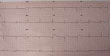
Left ventricular aneurysm can be associated with ST elevation.[1]
Causes
Ventricular aneurysms are usually complications resulting from a heart attack. When the heart muscle (cardiac muscle) partially dies during a heart attack, a layer of muscle may survive, and being severely weakened, start to become an aneurysm. The blood may flow into the surrounding dead muscle and inflate the weakened flap of muscle into a bubble. It may also be congenital.
Signs and symptoms
Ventricular aneurysms usually grow at a very slow pace, but can still pose problems. Usually this type of aneurysm grows in the left ventricle. This bubble has the potential to block blood flow to the body, and thus limit the patient's stamina. In other cases, a similarly developed pseudoaneurysm "false aneurysm" may burst, sometimes resulting in death of the patient. Also, blood clots may form on the inside of ventricular aneurysms, and form embolisms. If these blood clots escape from the aneurysm, they will be moved in the circulation throughout the body. If these blood clots get stuck inside a blood vessel, they may cause ischemia in a limb, a painful condition that can lead to reduced movement and tissue death in the limb. Alternatively, if they block a vessel going to the brain, they can cause a stroke. In certain cases, ventricular aneurysm may cause ventricular failure or arrythmia. At this stage, treatment is necessary.
Differential diagnosis
It should also not be confused with a pseudoaneurysm,[2][3] coronary artery aneurysm or a myocardial rupture (which involves a hole in the wall, not just a bulge.)
Investigations
When a person visits the hospital or doctor with other symptoms, especially with a history of heart problems, they will normally be required to undergo an electrocardiogram, which monitors electrical activity within the heart and shows abnormalities when a cardiac aneurysm is present. It can also appear as a bulge on a chest x-ray, and a more accurate diagnosis will then be made using an echocardiogram, which uses ultrasound to ‘photograph’ the heart and how it functions while it beats.
Treatment
Some people live with this type of aneurysm for many years without any specific treatment. Treatment is limited to surgery (ventricular reduction) for this defect of the heart. However, surgery is not required in most cases but, limiting the patient's physical activity levels to lower the risk of making the aneurysm bigger is advised. Also ACE Inhibitors seem to prevent Left Ventricular remodeling and aneurysm formation.
Blood thinning agents may be given to help reduce the likelihood of blood thickening and clots forming, along with the use of drugs to correct the irregular rhythm of the heart (seen on the electrocardiogram)
See also
- Atrial septal aneurysm
- Coronary artery aneurysm
References
- ↑ Victor F. Froelicher; Jonathan Myers (2006). Exercise and the heart. Elsevier Health Sciences. pp. 138–. ISBN 978-1-4160-0311-3. Retrieved 10 October 2010.
- ↑ Zoffoli G, Mangino D, Venturini A, et al. (February 2009). "Diagnosing left ventricular aneurysm from pseudo-aneurysm: a case report and a review in literature". J Cardiothorac Surg. 4 (1): 11. doi:10.1186/1749-8090-4-11. PMC 2654444
 . PMID 19239694.
 - ↑ Brown SL, Gropler RJ, Harris KM (May 1997). "Distinguishing left ventricular aneurysm from pseudoaneurysm. A review of the literature". Chest. 111 (5): 1403–9. doi:10.1378/chest.111.5.1403. PMID 9149600.
- Lohr, John T. "Ventricular Aneurysm: Treatment". Answers.com. Retrieved 22 September 2009.
- Lohr, John T. "Ventricular Aneurysm". Answers.com. Retrieved 22 September 2009.
- Graber, J.D.; Oakley, C.M.; Pickering, B.N.; Goodwin, J.F.; Raphael, M.J.; Steiner, R.E. "Ventricular aneurysm. An appraisal of diagnosis and surgical treatment.". British Heart Journal. PubMed. Retrieved 22 September 2009.