Uveitis
| Uveitis (Also known as Iridocyclitis) | |
|---|---|
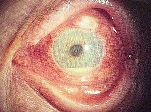 | |
| Hypopyon in anterior uveitis, seen as yellowish exudate in lower part of anterior chamber of eye | |
| Classification and external resources | |
| Specialty | Ophthalmology |
| ICD-10 | H20 |
| ICD-9-CM | 364 |
| DiseasesDB | 13676 |
| MedlinePlus | 001005 |
| eMedicine | oph/580 emerg/284 |
| Patient UK | Uveitis |
| MeSH | D014605 |
Uveitis is the inflammation of the uvea, the pigmented layer that lies between the inner retina and the outer fibrous layer composed of the sclera and cornea. The uvea consists of the middle layer of pigmented vascular structures of the eye and includes the iris, ciliary body, and choroid. Uveitis is an ophthalmic emergency and requires a thorough examination by an optometrist or ophthalmologist and urgent treatment to control the inflammation.
Classification
Uveitis is classified anatomically into anterior, intermediate, posterior, and panuveitic forms—based on the part of the eye primarily affected.[1] Prior to the twentieth century, uveitis was typically referred to in English as "ophthalmia."[2]
- Anterior uveitis includes iridocyclitis and iritis. Iritis is the inflammation of the anterior chamber and iris. Iridocyclitis presents the same symptoms as iritis, but also includes inflammation in the ciliary body.[3] Anywhere from two-thirds to 90% of uveitis cases are anterior in location. This condition can occur as a single episode and subside with proper treatment or may take on a recurrent or chronic nature.
- intermediate uveitis, also known as pars planitis, consists of vitritis—which is inflammation of cells in the vitreous cavity, sometimes with snowbanking, or deposition of inflammatory material on the pars plana. There are also "snowballs," which are inflammatory cells in the vitreous.
- Posterior uveitis or chorioretinitis is the inflammation of the retina and choroid.
- Pan-uveitis is the inflammation of all layers of the uvea.
Symptoms and signs
Anterior uveitis
- Burning of the eye
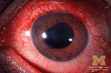
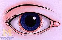
- Redness of the eye
- Blurred vision
- Photophobia or sensitivity to light
- Irregular pupil
- Eye pain
- Floaters, which are dark spots that float in the visual field
- Headaches
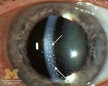
- Signs of anterior uveitis include dilated ciliary vessels, presence of cells and flare in the anterior chamber, and keratic precipitates ("KP") on the posterior surface of the cornea. In severe inflammation there may be evidence of a hypopyon. Old episodes of uveitis are identified by pigment deposits on lens, KPs, and festooned pupil on dilation of pupil.
- Busacca nodules, inflammatory nodules located on the surface of the iris in granulomatous forms of anterior uveitis such as Fuchs heterochromic iridocyclitis (FHI).[4]
- Synechia
Intermediate uveitis
Most common:
- Floaters
- Blurred vision
Intermediate uveitis normally only affects one eye. Less common is the presence of pain and photophobia.[5]
Posterior uveitis
Inflammation in the back of the eye is commonly characterized by:
- Floaters
- Blurred vision
- Photopsia or seeing flashing lights
Causes
Uveitis is usually an isolated illness, but can be associated with many other medical conditions.
In anterior uveitis, no associated condition or syndrome is found in approximately one-half of cases. However, anterior uveitis is often one of the syndromes associated with HLA-B27. Presence this type of HLA allele has a relative risk of evolving this disease by approximately 15%.[6]
The most common form of uveitis is acute anterior uveitis (AAU). It is most commonly associated with HLA-B27, which has important features: HLA-B27 AAU can be associated with ocular inflammation alone or in association with systemic disease. HLA-B27 AAU has characteristic clinical features including male preponderance, unilateral alternating acute onset, a non-granulomatous appearance, and frequent recurrences whereas HLA-B27 negative AAU has an equivalent male to female onset, bilateral chronic course, and more frequent granulomatous appearance.[7] Rheumatoid arthritis is not uncommon in Asian countries as a significant association of uveitis.[8]
Noninfectious or autoimmune causes
- Behçet disease
- Crohn's disease
- Fuchs heterochromic iridocyclitis
- Granulomatosis with polyangiitis
- HLA-B27 related uveitis
- Juvenile idiopathic arthritis
- Sarcoidosis
- Spondyloarthritis
- Sympathetic ophthalmia
- Tubulointerstitial nephritis and uveitis syndrome
Infectious causes
Uveitis may be an immune response to fight an infection inside the eye. While representing the minority of patients with uveitis, such possible infections include:
- brucellosis
- leptospirosis
- Lyme disease
- presumed ocular histoplasmosis syndrome
- syphilis
- toxocariasis
- toxoplasmic chorioretinitis
- tuberculosis
- Zika fever[9]
Associated with systemic diseases
Systemic disorders that can be associated with uveitis include:[10][11]
- ankylosing spondylitis
- Behçet's disease
- chronic granulomatous disease
- enthesitis
- inflammatory bowel disease
- juvenile rheumatoid arthritis
- Kawasaki's disease
- multiple sclerosis
- polyarteritis nodosa
- psoriatic arthritis
- reactive arthritis
- sarcoidosis
- systemic lupus erythematosus
- Vogt–Koyanagi–Harada disease
- Whipple's disease
Drug related side effects
- Rifabutin, a derivative of Rifampin has been shown to cause uveitis.[12]
- Several reports suggest the use of quinolones especially Moxifloxacin may lead to uveitis.[13]
- All of the widely [14] administered vaccines have been reported to cause uveitis.
White Dot syndromes
Occasionally, uveitis is not associated with a systemic condition: the inflammation is confined to the eye and has unknown etiology. In some of these cases, the presentation in the eye is characteristic of a described syndrome, which are called white dot syndromes, and include the following diagnoses:
- acute posterior multifocal placoid pigment epitheliopathy
- birdshot chorioretinopathy
- multifocal choroiditis and panuveitis
- multiple evanescent white dot syndrome
- punctate inner choroiditis
- serpiginous choroiditis
- acute zonal occult outer retinopathy
Masquerade syndromes
Masquerade syndromes are those conditions that include the presence of intraocular cells but are not due to immune-mediated uveitis entities. These may be divided into neoplastic and non-neoplastic conditions.
- Non-neoplastic:
- retinitis pigmentosa
- intraocular foreign body
- juvenile xanthogranuloma
- retinal detachment
- Neoplastic:
Pathophysiology
Immunologic factors
Onset of uveitis can broadly be described as a failure of the ocular immune system and the disease results from inflammation and tissue destruction. Uveitis is driven by the Th17 T cell sub-population that bear T-cell receptors specific for proteins found in the eye.[15] These are often not deleted centrally whether due to ocular antigen not being presented in the thymus (therefore not negatively selected) or a state of anergy is induced to prevent self targeting.[16][17]
Autoreactive T cells must normally be held in check by the suppressive environment produced by microglia and dendritic cells in the eye.[18] These cells produce large amounts of TGF beta and other suppressive cytokines, including IL-10, to prevent damage to the eye by reducing inflammation and causing T cells to differentiate to inducible T reg cells. Innate immune stimulation by bacteria and cellular stress is normally suppressed by myeloid suppression while inducible Treg cells prevent activation and clonal expansion of the autoreactive Th1 and Th17 cells that possess potential to cause damage to the eye.
Whether through infection or other causes, this balance can be upset and autoreactive T cells allowed to proliferate and migrate to the eye. Upon entry to the eye, these cells may be returned to an inducible Treg state by the presence of IL-10 and TGF-beta from microglia. Failure of this mechanism leads to neutrophil and other leukocyte recruitment from the peripheral blood through IL-17 secretion. Tissue destruction is mediated by non-specific macrophage activation and the resulting cytokine cascades.[19] Serum TNF-α is significantly elevated in cases while IL-6 and IL-8 are present in significantly higher quantities in the aqueous humour in patients with both quiescent and active uveitis.[20] These are inflammatory markers that non-specifically activate local macrophages causing tissue damage.
Genetic factors
The cause of non-infectious uveitis is unknown but there are some strong genetic factors that predispose disease onset including HLA-B27[21][22] and the PTPN22 genotype.[23]
Infectious agents
Recent evidence has pointed to reactivation of herpes simplex, varicella zoster and other viruses as important causes of developing what was previously described as idiopathic anterior uveitis.[24] Bacterial infection is another significant contributing factor in developing uveitis.[25]
Diagnosis
Diagnosis includes dilated fundus examination to rule out posterior uveitis, which presents with white spots across the retina along with retinitis and vasculitis.
Laboratory testing is usually used to diagnose specific underlying diseases, including rheumatologic tests (e.g. antinuclear antibody, rheumatoid factor, angiotensin converting enzyme inhibitor) and serology for infectious diseases (Syphilis, Toxoplasmosis, Tuberculosis).
Major histocompatibility antigen testing may be performed to investigate genetic susceptibility to uveitis. The most common antigens include HLA-B27, HLA-A29 (in birdshot chorioretinopathy) and HLA-B51 (in Behçet disease).
Radiology X-ray may be used to show coexisting arthritis and chest X-ray may be helpful in sarcoidosis.
Treatment
Uveitis is typically treated with glucocorticoid steroids, either as topical eye drops (prednisolone acetate) or as oral therapy.[26] Prior to the administration of corticosteroids, corneal ulcers must be ruled out. This is typically done using a fluoresence dye test.[27] In addition to corticosteroids, topical cycloplegics, such as atropine or homatropine, may be used. Successful treatment of active uveitis increases T-regulatory cells in the eye, which likely contributes to disease regression.[28] In some cases an injection of posterior subtenon triamcinolone acetate may also be given to reduce the swelling of the eye. [29]
Antimetabolite medications, such as methotrexate are often used for recalcitrant or more aggressive cases of uveitis. Experimental treatments with Infliximab or other anti-TNF infusions may prove helpful.
The anti-diabetic drug metformin is reported to inhibit the process that causes the inflammation in uveitis.[30]
In the case of herpetic uveitis, anti-viral medications, such as valaciclovir or aciclovir, may be administered to treat the causative viral infection.[31]
Prognosis
The prognosis is generally good for those who receive prompt diagnosis and treatment, but serious complication including cataracts, glaucoma, band keratopathy, macular edema and permanent vision loss may result if left untreated. The type of uveitis, as well as its severity, duration, and responsiveness to treatment or any associated illnesses, all factor into the outlook.[32]
Epidemiology
Uveitis affects approximately 1 in 4500 people and is most common between the ages 20 to 60 with men and women affected equally.
In western countries, anterior uveitis accounts for between 50% and 90% of uveitis cases. In Asian countries the proportion is between 28% and 50%.[33]
Uveitis is estimated to be responsible for approximately 10%-20% of the blindness in the United States.[34]
See also
- List of eye diseases and disorders
- List of systemic diseases with ocular manifestations
- Intermediate uveitis
References
- ↑ Jabs DA, Nussenblatt RB, Rosenbaum JT. Standardization of Uveitis Nomenclature (SUN) Working Group. Standardization of uveitis nomenclature for reporting clinical data. Results of the First International Workshop. Am J Ophthalmol 2005;140:509-516.
- ↑ Leffler CT, Schwartz SG, Stackhouse R, Davenport B, Spetzler K (2013). "Evolution and impact of eye and vision terms in written English". JAMA Ophthalmol. 131 (12): 1625–31. doi:10.1001/jamaophthalmol.2013.917. PMID 24337558.
- ↑ http://www.meduweb.com/showthread.php?t=29564
- ↑ Abdullah Al-Fawaz; Ralph D Levinson (25 Feb 2010). "Uveitis, Anterior, Granulomatous". eMedicine from WebMD. Retrieved 15 December 2010.
- ↑ Babu, BM; Rathinam, SR (Jan–Feb 2010). "Intermediate uveitis.". Indian journal of ophthalmology. 58 (1): 21–7. doi:10.4103/0301-4738.58469. PMC 2841370
 . PMID 20029143.
 - ↑ Table 5-7 in: Mitchell, Richard Sheppard; Kumar, Vinay; Abbas, Abul K.; Fausto, Nelson. Robbins Basic Pathology. Philadelphia: Saunders. ISBN 1-4160-2973-7. 8th edition.
- ↑ Larson, T; Nussenblatt, RB; Sen, HN (June 2011). "Emerging drugs for uveitis". Expert opinion on emerging drugs. 16 (2): 309–22. doi:10.1517/14728214.2011.537824. PMC 3102121. PMID 21210752.
- ↑ Imtiaz Ali Shah , Zuberi BF, Sangi SA, Abbasi SA: Systemic Manifestations of Iridocyclitis: Pak J Ophthalmol 1999 Vol. 15, No. 2; p,61-64.
- ↑ https://consumer.healthday.com/infectious-disease-information-21/virus-health-news-697/zika-infection-can-also-strike-eyes-of-adults-report-712182.html
- ↑ White G. "Uveitis." AllAboutVision.com. Retrieved August 20, 2006.
- ↑ McGonagle D, McDermott MF (2006) A proposed classification of the immunological diseases" PLoS Med 3(8) e297. doi:10.1371/journal.pmed.0030297
- ↑ CDC: Department of Human Services (9 September 1994). "Uveitis Associated with Rifabutin Therapy". 43(35);658: Morbidity and Mortality Weekly Report. Retrieved 5 May 2013.
- ↑ "Risk for Uveitis With Oral Moxifloxacin". JAMA Ophthalmology online. 2 October 2014.
- ↑ http://joii-journal.springeropen.com/articles/10.1186/1869-5760-3-43
- ↑ Nian, H.; Liang, D.; Zuo, A.; Wei, R.; Shao, H.; Born, W. K.; Kaplan, H. J.; Sun, D. (12 January 2012). "Characterization of Autoreactive and Bystander IL-17+ T Cells Induced in Immunized C57BL/6 Mice". Investigative Ophthalmology & Visual Science. 53 (2): 897–905. doi:10.1167/iovs.11-8297.
- ↑ Lambe T, Leung JC, Ferry H, et al. (April 2007). "Limited peripheral T cell anergy predisposes to retinal autoimmunity". J. Immunol. 178 (7): 4276–83. doi:10.4049/jimmunol.178.7.4276. PMID 17371984.
- ↑ Avichezer D, Grajewski RS, Chan CC, et al. (December 2003). "An immunologically privileged retinal antigen elicits tolerance: major role for central selection mechanisms". J. Exp. Med. 198 (11): 1665–76. doi:10.1084/jem.20030413. PMC 2194140. PMID 14657219.
- ↑ Forrester, JV; Xu, H; Kuffová, L; Dick, AD; McMenamin, PG (March 2010). "Dendritic cell physiology and function in the eye". Immunological reviews. 234 (1): 282–304. doi:10.1111/j.0105-2896.2009.00873.x. PMID 20193026.
- ↑ Khera, TK; Copland, DA; Boldison, J; Lait, PJ; Szymkowski, DE; Dick, AD; Nicholson, LB (May 2012). "Tumour necrosis factor-mediated macrophage activation in the target organ is critical for clinical manifestation of uveitis". Clinical and experimental immunology. 168 (2): 165–77. doi:10.1111/j.1365-2249.2012.04567.x. PMC 3390517. PMID 22471277.
- ↑ Valentincic, NV; de Groot-Mijnes, JD; Kraut, A; Korosec, P; Hawlina, M; Rothova, A; Kraut, A; Korosec, P; Hawlina, M; Rothova, A (2011). "Intraocular and serum cytokine profiles in patients with intermediate uveitis". Molecular vision. 17: 2003–10. PMC 3154134. PMID 21850175.
- ↑ Wakefield, D; Chang, JH; Amjadi, S; Maconochie, Z; Abu El-Asrar, A; McCluskey, P (April 2011). "What is new HLA-B27 acute anterior uveitis?". Ocular immunology and inflammation. 19 (2): 139–44. doi:10.3109/09273948.2010.542269. PMID 21428757.
- ↑ Caspi, Rachel R. (2010). "A look at autoimmunity and inflammation in the eye". Journal of Clinical Investigation. 120 (9): 3073–3083. doi:10.1172/JCI42440. PMC 2929721. PMID 20811163.
- ↑ Burn, Garth L.; Svensson, Lena; Sanchez-Blanco, Cristina; Saini, Manoj; Cope, Andrew P. (1 December 2011). "Why is PTPN22 a good candidate susceptibility gene for autoimmune disease?". FEBS Letters. 585 (23): 3689–3698. doi:10.1016/j.febslet.2011.04.032. PMID 21515266.
- ↑ Jap, A; Chee, SP (November 2011). "Viral anterior uveitis". Current opinion in ophthalmology. 22 (6): 483–8. doi:10.1097/ICU.0b013e32834be021. PMID 21918442.
- ↑ Dick, Andrew D. (1 January 2012). "Road to Fulfilment: Taming the Immune Response to Restore Vision". Ophthalmic Research. 48 (1): 43–49. doi:10.1159/000335982. PMID 22398563.
- ↑ Pato, E; Muñoz-Fernández, S; Francisco, F; Abad, MA; Maese, J; Ortiz, A; Carmona, L; Uveitis Working Group from Spanish Society of, Rheumatology (February 2011). "Systematic review on the effectiveness of immunosuppressants and biological therapies in the treatment of autoimmune posterior uveitis". Seminars in arthritis and rheumatism. 40 (4): 314–23. doi:10.1016/j.semarthrit.2010.05.008. PMID 20656330.
- ↑ "Fluorescein eye stain". NIH. Retrieved 15 May 2012.
- ↑ Ruggieri, S; Frassanito, MA; Dammacco, R; Guerriero, S (2012-05-07). "Treg Lymphocytes in Autoimmune Uveitis". Ocular immunology and inflammation. 20 (4): 255–61. doi:10.3109/09273948.2012.681830. PMID 22564107.
- ↑ BNF 45 March 2003
- ↑ "Diabetes drug could treat leading cause of blindness". May 8, 2012.
- ↑ https://www.inkling.com/read/albert-jakobiecs-principles-practice-ophthalmology-3rd/chapter-49/herpetic-disease-of-the
- ↑ "Uveitis from intelihealth".
- ↑ Chang, JH; Wakefield, D (December 2002). "Uveitis: a global perspective". Ocular immunology and inflammation. 10 (4): 263–79. doi:10.1076/ocii.10.4.263.15592. PMID 12854035.
- ↑ Gritz, D; Wong, IG (1 March 2004). "Incidence and prevalence of uveitis in Northern California The Northern California Epidemiology of Uveitis Study". Ophthalmology. 111 (3): 491–500. doi:10.1016/j.ophtha.2003.06.014. PMID 15019324.
External links
- The Ocular Immunology and Uveitis Foundation Dedicated to research, education and support for patients with uveitis