Virus
| Viruses | |
|---|---|
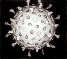 | |
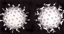
| Rotavirus | |
| Virus classification | |
| Group: | I–VII |
| Groups | |
|
I: dsDNA viruses | |
A virus is a small infectious agent that replicates only inside the living cells of other organisms. Viruses can infect all types of life forms, from animals and plants to microorganisms, including bacteria and archaea.[1]
Since Dmitri Ivanovsky's 1892 article describing a non-bacterial pathogen infecting tobacco plants, and the discovery of the tobacco mosaic virus by Martinus Beijerinck in 1898,[2] about 5,000 virus species have been described in detail,[3] although there are millions of types.[4] Viruses are found in almost every ecosystem on Earth and are the most abundant type of biological entity.[5][6] The study of viruses is known as virology, a sub-speciality of microbiology.
While not inside an infected cell or in the process of infecting a cell, viruses exist in the form of independent particles. These viral particles, also known as virions, consist of two or three parts: (i) the genetic material made from either DNA or RNA, long molecules that carry genetic information; (ii) a protein coat, called the capsid, which surrounds and protects the genetic material; and in some cases (iii) an envelope of lipids that surrounds the protein coat when they are outside a cell. The shapes of these virus particles range from simple helical and icosahedral forms for some virus species to more complex structures for others. Most virus species have virions that are too small to be seen with an optical microscope. The average virion is about one one-hundredth the size of the average bacterium.
The origins of viruses in the evolutionary history of life are unclear: some may have evolved from plasmids—pieces of DNA that can move between cells—while others may have evolved from bacteria. In evolution, viruses are an important means of horizontal gene transfer, which increases genetic diversity.[7] Viruses are considered by some to be a life form, because they carry genetic material, reproduce, and evolve through natural selection. However they lack key characteristics (such as cell structure) that are generally considered necessary to count as life. Because they possess some but not all such qualities, viruses have been described as "organisms at the edge of life",[8] and as replicators.[9]
Viruses spread in many ways; viruses in plants are often transmitted from plant to plant by insects that feed on plant sap, such as aphids; viruses in animals can be carried by blood-sucking insects. These disease-bearing organisms are known as vectors. Influenza viruses are spread by coughing and sneezing. Norovirus and rotavirus, common causes of viral gastroenteritis, are transmitted by the faecal–oral route and are passed from person to person by contact, entering the body in food or water. HIV is one of several viruses transmitted through sexual contact and by exposure to infected blood. The range of host cells that a virus can infect is called its "host range". This can be narrow, meaning a virus is capable of infecting few species, or broad, meaning it is capable of infecting many.[10]
Viral infections in animals provoke an immune response that usually eliminates the infecting virus. Immune responses can also be produced by vaccines, which confer an artificially acquired immunity to the specific viral infection. However, some viruses including those that cause AIDS and viral hepatitis evade these immune responses and result in chronic infections. Antibiotics have no effect on viruses, but several antiviral drugs have been developed.
Etymology
The word is from the Latin neuter vīrus referring to poison and other noxious liquids, from 'the same Indo-European base as Sanskrit viṣa poison, Avestan vīša poison, ancient Greek ἰός poison', first attested in English in 1398 in John Trevisa's translation of Bartholomeus Anglicus's De Proprietatibus Rerum.[11][12] Virulent, from Latin virulentus (poisonous), dates to c. 1400.[13][14] A meaning of "agent that causes infectious disease" is first recorded in 1728,[12] before the discovery of viruses by Dmitri Ivanovsky in 1892. The English plural is viruses (sometimes also viri[15] or vira[16]), whereas the Latin word is a mass noun, which has no classically attested plural (however in Neo-Latin vīra[17][18] is used). The adjective viral dates to 1948.[19] The term virion (plural virions), which dates from 1959,[20] is also used to refer to a single, stable infective viral particle that is released from the cell and is fully capable of infecting other cells of the same type.[21]
History
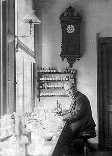
Louis Pasteur was unable to find a causative agent for rabies and speculated about a pathogen too small to be detected using a microscope.[22] In 1884, the French microbiologist Charles Chamberland invented a filter (known today as the Chamberland filter or the Pasteur-Chamberland filter) with pores smaller than bacteria. Thus, he could pass a solution containing bacteria through the filter and completely remove them from the solution.[23] In 1892, the Russian biologist Dmitri Ivanovsky used this filter to study what is now known as the tobacco mosaic virus. His experiments showed that crushed leaf extracts from infected tobacco plants remain infectious after filtration. Ivanovsky suggested the infection might be caused by a toxin produced by bacteria, but did not pursue the idea.[24] At the time it was thought that all infectious agents could be retained by filters and grown on a nutrient medium – this was part of the germ theory of disease.[2] In 1898, the Dutch microbiologist Martinus Beijerinck repeated the experiments and became convinced that the filtered solution contained a new form of infectious agent.[25] He observed that the agent multiplied only in cells that were dividing, but as his experiments did not show that it was made of particles, he called it a contagium vivum fluidum (soluble living germ) and re-introduced the word virus.[24] Beijerinck maintained that viruses were liquid in nature, a theory later discredited by Wendell Stanley, who proved they were particulate.[24] In the same year Friedrich Loeffler and Paul Frosch passed the first animal virus – agent of foot-and-mouth disease (aphthovirus) – through a similar filter.[26]
In the early 20th century, the English bacteriologist Frederick Twort discovered a group of viruses that infect bacteria, now called bacteriophages[27] (or commonly phages), and the French-Canadian microbiologist Félix d'Herelle described viruses that, when added to bacteria on an agar plate, would produce areas of dead bacteria. He accurately diluted a suspension of these viruses and discovered that the highest dilutions (lowest virus concentrations), rather than killing all the bacteria, formed discrete areas of dead organisms. Counting these areas and multiplying by the dilution factor allowed him to calculate the number of viruses in the original suspension.[28] Phages were heralded as a potential treatment for diseases such as typhoid and cholera, but their promise was forgotten with the development of penicillin. The study of phages provided insights into the switching on and off of genes, and a useful mechanism for introducing foreign genes into bacteria.
By the end of the 19th century, viruses were defined in terms of their infectivity, their ability to be filtered, and their requirement for living hosts. Viruses had been grown only in plants and animals. In 1906, Ross Granville Harrison invented a method for growing tissue in lymph, and, in 1913, E. Steinhardt, C. Israeli, and R. A. Lambert used this method to grow vaccinia virus in fragments of guinea pig corneal tissue.[29] In 1928, H. B. Maitland and M. C. Maitland grew vaccinia virus in suspensions of minced hens' kidneys. Their method was not widely adopted until the 1950s, when poliovirus was grown on a large scale for vaccine production.[30]
Another breakthrough came in 1931, when the American pathologist Ernest William Goodpasture and Alice Miles Woodruff grew influenza and several other viruses in fertilised chickens' eggs.[31] In 1949, John Franklin Enders, Thomas Weller, and Frederick Robbins grew polio virus in cultured human embryo cells, the first virus to be grown without using solid animal tissue or eggs. This work enabled Jonas Salk to make an effective polio vaccine.[32]
The first images of viruses were obtained upon the invention of electron microscopy in 1931 by the German engineers Ernst Ruska and Max Knoll.[33] In 1935, American biochemist and virologist Wendell Meredith Stanley examined the tobacco mosaic virus and found it was mostly made of protein.[34] A short time later, this virus was separated into protein and RNA parts.[35] The tobacco mosaic virus was the first to be crystallised and its structure could therefore be elucidated in detail. The first X-ray diffraction pictures of the crystallised virus were obtained by Bernal and Fankuchen in 1941. On the basis of her pictures, Rosalind Franklin discovered the full structure of the virus in 1955.[36] In the same year, Heinz Fraenkel-Conrat and Robley Williams showed that purified tobacco mosaic virus RNA and its protein coat can assemble by themselves to form functional viruses, suggesting that this simple mechanism was probably the means through which viruses were created within their host cells.[37]
The second half of the 20th century was the golden age of virus discovery and most of the over 2,000 recognised species of animal, plant, and bacterial viruses were discovered during these years.[38] In 1957, equine arterivirus and the cause of Bovine virus diarrhoea (a pestivirus) were discovered. In 1963, the hepatitis B virus was discovered by Baruch Blumberg,[39] and in 1965, Howard Temin described the first retrovirus. Reverse transcriptase, the enzyme that retroviruses use to make DNA copies of their RNA, was first described in 1970, independently by Howard Martin Temin and David Baltimore.[40] In 1983 Luc Montagnier's team at the Pasteur Institute in France, first isolated the retrovirus now called HIV.[41] In 1989 Michael Houghton's team at Chiron Corporation discovered Hepatitis C.[42][43]
Origins
Viruses are found wherever there is life and have probably existed since living cells first evolved.[44] The origin of viruses is unclear because they do not form fossils, so molecular techniques have been used to compare the DNA or RNA of viruses and are a useful means of investigating how they arose.[45] In addition, viral genetic material may occasionally integrate into the germline of the host organisms, by which they can be passed on vertically to the offspring of the host for many generations. This provides an invaluable source of information for paleovirologists to trace back ancient viruses that have existed up to millions of years ago. There are three main hypotheses that aim to explain the origins of viruses:[46][47]
- Regressive hypothesis
- Viruses may have once been small cells that parasitised larger cells. Over time, genes not required by their parasitism were lost. The bacteria rickettsia and chlamydia are living cells that, like viruses, can reproduce only inside host cells. They lend support to this hypothesis, as their dependence on parasitism is likely to have caused the loss of genes that enabled them to survive outside a cell. This is also called the degeneracy hypothesis,[48][49] or reduction hypothesis.[50]
- Cellular origin hypothesis
- Some viruses may have evolved from bits of DNA or RNA that "escaped" from the genes of a larger organism. The escaped DNA could have come from plasmids (pieces of naked DNA that can move between cells) or transposons (molecules of DNA that replicate and move around to different positions within the genes of the cell).[51] Once called "jumping genes", transposons are examples of mobile genetic elements and could be the origin of some viruses. They were discovered in maize by Barbara McClintock in 1950.[52] This is sometimes called the vagrancy hypothesis,[48][53] or the escape hypothesis.[50]
- Co-evolution hypothesis
- This is also called the virus-first hypothesis[50] and proposes that viruses may have evolved from complex molecules of protein and nucleic acid at the same time as cells first appeared on Earth and would have been dependent on cellular life for billions of years. Viroids are molecules of RNA that are not classified as viruses because they lack a protein coat. However, they have characteristics that are common to several viruses and are often called subviral agents.[54] Viroids are important pathogens of plants.[55] They do not code for proteins but interact with the host cell and use the host machinery for their replication.[56] The hepatitis delta virus of humans has an RNA genome similar to viroids but has a protein coat derived from hepatitis B virus and cannot produce one of its own. It is, therefore, a defective virus. Although hepatitis delta virus genome may replicate independently once inside a host cell, it requires the help of hepatitis B virus to provide a protein coat so that it can be transmitted to new cells.[57] In similar manner, the sputnik virophage is dependent on mimivirus, which infects the protozoan Acanthamoeba castellanii.[58] These viruses, which are dependent on the presence of other virus species in the host cell, are called satellites and may represent evolutionary intermediates of viroids and viruses.[59][60]
In the past, there were problems with all of these hypotheses: the regressive hypothesis did not explain why even the smallest of cellular parasites do not resemble viruses in any way. The escape hypothesis did not explain the complex capsids and other structures on virus particles. The virus-first hypothesis contravened the definition of viruses in that they require host cells.[50] Viruses are now recognised as ancient and as having origins that pre-date the divergence of life into the three domains.[61] This discovery has led modern virologists to reconsider and re-evaluate these three classical hypotheses.[61]
The evidence for an ancestral world of RNA cells[62] and computer analysis of viral and host DNA sequences are giving a better understanding of the evolutionary relationships between different viruses and may help identify the ancestors of modern viruses. To date, such analyses have not proved which of these hypotheses is correct.[62] However, it seems unlikely that all currently known viruses have a common ancestor, and viruses have probably arisen numerous times in the past by one or more mechanisms.[63]
Prions are infectious protein molecules that do not contain DNA or RNA.[64] They can cause infections such as scrapie in sheep, bovine spongiform encephalopathy ("mad cow" disease) in cattle, and chronic wasting disease in deer; in humans, prionic diseases include Kuru, Creutzfeldt–Jakob disease, and Gerstmann–Sträussler–Scheinker syndrome.[65] Although prions are fundamentally different from viruses and viroids, their discovery gives credence to the theory that viruses could have evolved from self-replicating molecules.[66]
Microbiology
Life properties
Opinions differ on whether viruses are a form of life, or organic structures that interact with living organisms.[67] They have been described as "organisms at the edge of life",[8] since they resemble organisms in that they possess genes, evolve by natural selection,[68] and reproduce by creating multiple copies of themselves through self-assembly. Although they have genes, they do not have a cellular structure, which is often seen as the basic unit of life. Viruses do not have their own metabolism, and require a host cell to make new products. They therefore cannot naturally reproduce outside a host cell[69] – although bacterial species such as rickettsia and chlamydia are considered living organisms despite the same limitation.[70][71] Accepted forms of life use cell division to reproduce, whereas viruses spontaneously assemble within cells. They differ from autonomous growth of crystals as they inherit genetic mutations while being subject to natural selection. Virus self-assembly within host cells has implications for the study of the origin of life, as it lends further credence to the hypothesis that life could have started as self-assembling organic molecules.[1]
Structure
_Virus_PHIL_1878_lores.jpg)
Viruses display a wide diversity of shapes and sizes, called morphologies. In general, viruses are much smaller than bacteria. Most viruses that have been studied have a diameter between 20 and 300 nanometres. Some filoviruses have a total length of up to 1400 nm; their diameters are only about 80 nm.[72] Most viruses cannot be seen with an optical microscope so scanning and transmission electron microscopes are used to visualise virions.[73] To increase the contrast between viruses and the background, electron-dense "stains" are used. These are solutions of salts of heavy metals, such as tungsten, that scatter the electrons from regions covered with the stain. When virions are coated with stain (positive staining), fine detail is obscured. Negative staining overcomes this problem by staining the background only.[74]
A complete virus particle, known as a virion, consists of nucleic acid surrounded by a protective coat of protein called a capsid. These are formed from identical protein subunits called capsomeres.[75] Viruses can have a lipid "envelope" derived from the host cell membrane. The capsid is made from proteins encoded by the viral genome and its shape serves as the basis for morphological distinction.[76][77] Virally coded protein subunits will self-assemble to form a capsid, in general requiring the presence of the virus genome. Complex viruses code for proteins that assist in the construction of their capsid. Proteins associated with nucleic acid are known as nucleoproteins, and the association of viral capsid proteins with viral nucleic acid is called a nucleocapsid. The capsid and entire virus structure can be mechanically (physically) probed through atomic force microscopy.[78][79] In general, there are four main morphological virus types:
- Helical
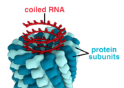
- These viruses are composed of a single type of capsomere stacked around a central axis to form a helical structure, which may have a central cavity, or tube. This arrangement results in rod-shaped or filamentous virions: These can be short and highly rigid, or long and very flexible. The genetic material, in general, single-stranded RNA, but ssDNA in some cases, is bound into the protein helix by interactions between the negatively charged nucleic acid and positive charges on the protein. Overall, the length of a helical capsid is related to the length of the nucleic acid contained within it and the diameter is dependent on the size and arrangement of capsomeres. The well-studied tobacco mosaic virus is an example of a helical virus.[80]
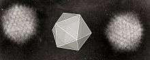
- Icosahedral
- Most animal viruses are icosahedral or near-spherical with chiral icosahedral symmetry. A regular icosahedron is the optimum way of forming a closed shell from identical sub-units. The minimum number of identical capsomeres required is twelve, each composed of five identical sub-units. Many viruses, such as rotavirus, have more than twelve capsomers and appear spherical but they retain this symmetry. Capsomeres at the apices are surrounded by five other capsomeres and are called pentons. Capsomeres on the triangular faces are surrounded by six others and are called hexons.[81] Hexons are in essence flat and pentons, which form the 12 vertices, are curved. The same protein may act as the subunit of both the pentamers and hexamers or they may be composed of different proteins.
- Prolate
- This is an icosahedron elongated along the fivefold axis and is a common arrangement of the heads of bacteriophages. This structure is composed of a cylinder with a cap at either end.[82]
- Envelope
- Some species of virus envelop themselves in a modified form of one of the cell membranes, either the outer membrane surrounding an infected host cell or internal membranes such as nuclear membrane or endoplasmic reticulum, thus gaining an outer lipid bilayer known as a viral envelope. This membrane is studded with proteins coded for by the viral genome and host genome; the lipid membrane itself and any carbohydrates present originate entirely from the host. The influenza virus and HIV use this strategy. Most enveloped viruses are dependent on the envelope for their infectivity.[83]
- Complex
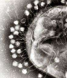
- These viruses possess a capsid that is neither purely helical nor purely icosahedral, and that may possess extra structures such as protein tails or a complex outer wall. Some bacteriophages, such as Enterobacteria phage T4, have a complex structure consisting of an icosahedral head bound to a helical tail, which may have a hexagonal base plate with protruding protein tail fibres. This tail structure acts like a molecular syringe, attaching to the bacterial host and then injecting the viral genome into the cell.[84]
The poxviruses are large, complex viruses that have an unusual morphology. The viral genome is associated with proteins within a central disc structure known as a nucleoid. The nucleoid is surrounded by a membrane and two lateral bodies of unknown function. The virus has an outer envelope with a thick layer of protein studded over its surface. The whole virion is slightly pleiomorphic, ranging from ovoid to brick shape.[85] Mimivirus is one of the largest characterised viruses, with a capsid diameter of 400 nm. Protein filaments measuring 100 nm project from the surface. The capsid appears hexagonal under an electron microscope, therefore the capsid is probably icosahedral.[86] In 2011, researchers discovered the largest then known virus in samples of water collected from the ocean floor off the coast of Las Cruces, Chile. Provisionally named Megavirus chilensis, it can be seen with a basic optical microscope.[87] In 2013, the Pandoravirus genus was discovered in Chile and Australia, and has genomes about twice as large as Megavirus and Mimivirus.[88]
Some viruses that infect Archaea have complex structures that are unrelated to any other form of virus, with a wide variety of unusual shapes, ranging from spindle-shaped structures, to viruses that resemble hooked rods, teardrops or even bottles. Other archaeal viruses resemble the tailed bacteriophages, and can have multiple tail structures.[89]
Genome
| Property | Parameters |
|---|---|
| Nucleic acid |
|
| Shape |
|
| Strandedness |
|
| Sense |
|
An enormous variety of genomic structures can be seen among viral species; as a group, they contain more structural genomic diversity than plants, animals, archaea, or bacteria. There are millions of different types of viruses,[4] although only about 5,000 types have been described in detail.[3] As of September 2015, the NCBI Virus genome database has more than 75,000 complete genome sequences.[90] but there are doubtlessly many more to be discovered.[91][92]
A virus has either a DNA or an RNA genome and is called a DNA virus or an RNA virus, respectively. The vast majority of viruses have RNA genomes. Plant viruses tend to have single-stranded RNA genomes and bacteriophages tend to have double-stranded DNA genomes.[93]
Viral genomes are circular, as in the polyomaviruses, or linear, as in the adenoviruses. The type of nucleic acid is irrelevant to the shape of the genome. Among RNA viruses and certain DNA viruses, the genome is often divided up into separate parts, in which case it is called segmented. For RNA viruses, each segment often codes for only one protein and they are usually found together in one capsid. However, all segments are not required to be in the same virion for the virus to be infectious, as demonstrated by brome mosaic virus and several other plant viruses.[72]
A viral genome, irrespective of nucleic acid type, is almost always either single-stranded or double-stranded. Single-stranded genomes consist of an unpaired nucleic acid, analogous to one-half of a ladder split down the middle. Double-stranded genomes consist of two complementary paired nucleic acids, analogous to a ladder. The virus particles of some virus families, such as those belonging to the Hepadnaviridae, contain a genome that is partially double-stranded and partially single-stranded.[93]
For most viruses with RNA genomes and some with single-stranded DNA genomes, the single strands are said to be either positive-sense (called the plus-strand) or negative-sense (called the minus-strand), depending on if they are complementary to the viral messenger RNA (mRNA). Positive-sense viral RNA is in the same sense as viral mRNA and thus at least a part of it can be immediately translated by the host cell. Negative-sense viral RNA is complementary to mRNA and thus must be converted to positive-sense RNA by an RNA-dependent RNA polymerase before translation. DNA nomenclature for viruses with single-sense genomic ssDNA is similar to RNA nomenclature, in that the template strand for the viral mRNA is complementary to it (−), and the coding strand is a copy of it (+).[93] However, several types of ssDNA and ssRNA viruses have genomes that are ambisense in that transcription can occur off both strands in a double-stranded replicative intermediate. Examples include geminiviruses, which are ssDNA plant viruses and arenaviruses, which are ssRNA viruses of animals.[94]
Genome size varies greatly between species. The smallest viral genomes – the ssDNA circoviruses, family Circoviridae – code for only two proteins and have a genome size of only two kilobases;[95] the largest–the pandoraviruses–have genome sizes of around two megabases which code for about 2500 proteins.[96]
In general, RNA viruses have smaller genome sizes than DNA viruses because of a higher error-rate when replicating, and have a maximum upper size limit.[45] Beyond this limit, errors in the genome when replicating render the virus useless or uncompetitive. To compensate for this, RNA viruses often have segmented genomes – the genome is split into smaller molecules – thus reducing the chance that an error in a single-component genome will incapacitate the entire genome. In contrast, DNA viruses generally have larger genomes because of the high fidelity of their replication enzymes.[97] Single-strand DNA viruses are an exception to this rule, however, as mutation rates for these genomes can approach the extreme of the ssRNA virus case.[98]
Genetic mutation
Viruses undergo genetic change by several mechanisms. These include a process called antigenic drift where individual bases in the DNA or RNA mutate to other bases. Most of these point mutations are "silent" – they do not change the protein that the gene encodes – but others can confer evolutionary advantages such as resistance to antiviral drugs.[99][100] Antigenic shift occurs when there is a major change in the genome of the virus. This can be a result of recombination or reassortment. When this happens with influenza viruses, pandemics might result.[101] RNA viruses often exist as quasispecies or swarms of viruses of the same species but with slightly different genome nucleoside sequences. Such quasispecies are a prime target for natural selection.[102]
Segmented genomes confer evolutionary advantages; different strains of a virus with a segmented genome can shuffle and combine genes and produce progeny viruses or (offspring) that have unique characteristics. This is called reassortment or viral sex.[103]
Genetic recombination is the process by which a strand of DNA is broken and then joined to the end of a different DNA molecule. This can occur when viruses infect cells simultaneously and studies of viral evolution have shown that recombination has been rampant in the species studied.[104] Recombination is common to both RNA and DNA viruses.[105][106]
Replication cycle
Viral populations do not grow through cell division, because they are acellular. Instead, they use the machinery and metabolism of a host cell to produce multiple copies of themselves, and they assemble in the cell.

The life cycle of viruses differs greatly between species but there are six basic stages in the life cycle of viruses:[107]
Attachment is a specific binding between viral capsid proteins and specific receptors on the host cellular surface. This specificity determines the host range of a virus. For example, HIV infects a limited range of human leucocytes. This is because its surface protein, gp120, specifically interacts with the CD4 molecule – a chemokine receptor – which is most commonly found on the surface of CD4+ T-Cells. This mechanism has evolved to favour those viruses that infect only cells in which they are capable of replication. Attachment to the receptor can induce the viral envelope protein to undergo changes that results in the fusion of viral and cellular membranes, or changes of non-enveloped virus surface proteins that allow the virus to enter.
Penetration follows attachment: Virions enter the host cell through receptor-mediated endocytosis or membrane fusion. This is often called viral entry. The infection of plant and fungal cells is different from that of animal cells. Plants have a rigid cell wall made of cellulose, and fungi one of chitin, so most viruses can get inside these cells only after trauma to the cell wall.[108] However, nearly all plant viruses (such as tobacco mosaic virus) can also move directly from cell to cell, in the form of single-stranded nucleoprotein complexes, through pores called plasmodesmata.[109] Bacteria, like plants, have strong cell walls that a virus must breach to infect the cell. However, given that bacterial cell walls are much less thick than plant cell walls due to their much smaller size, some viruses have evolved mechanisms that inject their genome into the bacterial cell across the cell wall, while the viral capsid remains outside.[110]
Uncoating is a process in which the viral capsid is removed: This may be by degradation by viral enzymes or host enzymes or by simple dissociation; the end-result is the releasing of the viral genomic nucleic acid.
Replication of viruses involves primarily multiplication of the genome. Replication involves synthesis of viral messenger RNA (mRNA) from "early" genes (with exceptions for positive sense RNA viruses), viral protein synthesis, possible assembly of viral proteins, then viral genome replication mediated by early or regulatory protein expression. This may be followed, for complex viruses with larger genomes, by one or more further rounds of mRNA synthesis: "late" gene expression is, in general, of structural or virion proteins.
Assembly – Following the structure-mediated self-assembly of the virus particles, some modification of the proteins often occurs. In viruses such as HIV, this modification (sometimes called maturation) occurs after the virus has been released from the host cell.[111]
Release – Viruses can be released from the host cell by lysis, a process that kills the cell by bursting its membrane and cell wall if present: This is a feature of many bacterial and some animal viruses. Some viruses undergo a lysogenic cycle where the viral genome is incorporated by genetic recombination into a specific place in the host's chromosome. The viral genome is then known as a "provirus" or, in the case of bacteriophages a "prophage".[112] Whenever the host divides, the viral genome is also replicated. The viral genome is mostly silent within the host. However, at some point, the provirus or prophage may give rise to active virus, which may lyse the host cells.[113] Enveloped viruses (e.g., HIV) typically are released from the host cell by budding. During this process the virus acquires its envelope, which is a modified piece of the host's plasma or other, internal membrane.[114]
The genetic material within virus particles, and the method by which the material is replicated, varies considerably between different types of viruses.
- DNA viruses
- The genome replication of most DNA viruses takes place in the cell's nucleus. If the cell has the appropriate receptor on its surface, these viruses enter the cell sometimes by direct fusion with the cell membrane (e.g., herpesviruses) or – more usually – by receptor-mediated endocytosis. Most DNA viruses are entirely dependent on the host cell's DNA and RNA synthesising machinery, and RNA processing machinery. However, viruses with larger genomes may encode much of this machinery themselves. In eukaryotes the viral genome must cross the cell's nuclear membrane to access this machinery, while in bacteria it need only enter the cell.[115]
- RNA viruses
- Replication usually takes place in the cytoplasm. RNA viruses can be placed into four different groups depending on their modes of replication. The polarity (whether or not it can be used directly by ribosomes to make proteins) of single-stranded RNA viruses largely determines the replicative mechanism; the other major criterion is whether the genetic material is single-stranded or double-stranded. All RNA viruses use their own RNA replicase enzymes to create copies of their genomes.[116]
- Reverse transcribing viruses
- These have ssRNA (Retroviridae, Metaviridae, Pseudoviridae) or dsDNA (Caulimoviridae, and Hepadnaviridae) in their particles. Reverse transcribing viruses with RNA genomes (retroviruses), use a DNA intermediate to replicate, whereas those with DNA genomes (pararetroviruses) use an RNA intermediate during genome replication. Both types use a reverse transcriptase, or RNA-dependent DNA polymerase enzyme, to carry out the nucleic acid conversion. Retroviruses integrate the DNA produced by reverse transcription into the host genome as a provirus as a part of the replication process; pararetroviruses do not, although integrated genome copies of especially plant pararetroviruses can give rise to infectious virus.[117] They are susceptible to antiviral drugs that inhibit the reverse transcriptase enzyme, e.g. zidovudine and lamivudine. An example of the first type is HIV, which is a retrovirus. Examples of the second type are the Hepadnaviridae, which includes Hepatitis B virus.[118]
Effects on the host cell
The range of structural and biochemical effects that viruses have on the host cell is extensive.[119] These are called cytopathic effects.[120] Most virus infections eventually result in the death of the host cell. The causes of death include cell lysis, alterations to the cell's surface membrane and apoptosis.[121] Often cell death is caused by cessation of its normal activities because of suppression by virus-specific proteins, not all of which are components of the virus particle.[122]
Some viruses cause no apparent changes to the infected cell. Cells in which the virus is latent and inactive show few signs of infection and often function normally.[123] This causes persistent infections and the virus is often dormant for many months or years. This is often the case with herpes viruses.[124][125] Some viruses, such as Epstein–Barr virus, can cause cells to proliferate without causing malignancy,[126] while others, such as papillomaviruses, are established causes of cancer.[127]
Host range
Viruses are by far the most abundant biological entities on Earth and they outnumber all the others put together.[128] They infect all types of cellular life including animals, plants, bacteria and fungi.[3] However, different types of viruses can infect only a limited range of hosts and many are species-specific. Some, such as smallpox virus for example, can infect only one species – in this case humans,[129] and are said to have a narrow host range. Other viruses, such as rabies virus, can infect different species of mammals and are said to have a broad range.[130] The viruses that infect plants are harmless to animals, and most viruses that infect other animals are harmless to humans.[131] The host range of some bacteriophages is limited to a single strain of bacteria and they can be used to trace the source of outbreaks of infections by a method called phage typing.[132]
Classification
Classification seeks to describe the diversity of viruses by naming and grouping them on the basis of similarities. In 1962, André Lwoff, Robert Horne, and Paul Tournier were the first to develop a means of virus classification, based on the Linnaean hierarchical system.[133] This system bases classification on phylum, class, order, family, genus, and species. Viruses were grouped according to their shared properties (not those of their hosts) and the type of nucleic acid forming their genomes.[134] Later the International Committee on Taxonomy of Viruses was formed. However, viruses are not classified on the basis of phylum or class, as their small genome size and high rate of mutation makes it difficult to determine their ancestry beyond order. As such, the Baltimore classification is used to supplement the more traditional hierarchy.
ICTV classification
The International Committee on Taxonomy of Viruses (ICTV) developed the current classification system and wrote guidelines that put a greater weight on certain virus properties to maintain family uniformity. A unified taxonomy (a universal system for classifying viruses) has been established. The 9th lCTV Report[135] defines the concept of the virus species as the lowest taxon (group) in a branching hierarchy of viral taxa.[136] However, at present only a small part of the total diversity of viruses has been studied, with analyses of samples from humans finding that about 20% of the virus sequences recovered have not been seen before, and samples from the environment, such as from seawater and ocean sediments, finding that the large majority of sequences are completely novel.[137]
The general taxonomic structure is as follows:
In the current (2013) ICTV taxonomy, 7 orders have been established, the Caudovirales, Herpesvirales, Ligamenvirales, Mononegavirales, Nidovirales, Picornavirales, and Tymovirales. The committee does not formally distinguish between subspecies, strains, and isolates. In total there are 7 orders, 103 families, 22 subfamilies, 455 genera, about 2,827 species and over 4,000 types yet unclassified.[135][138][139]
Baltimore classification
The Nobel Prize-winning biologist David Baltimore devised the Baltimore classification system.[40][140] The ICTV classification system is used in conjunction with the Baltimore classification system in modern virus classification.[141][142][143]
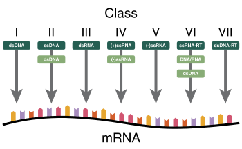
The Baltimore classification of viruses is based on the mechanism of mRNA production. Viruses must generate mRNAs from their genomes to produce proteins and replicate themselves, but different mechanisms are used to achieve this in each virus family. Viral genomes may be single-stranded (ss) or double-stranded (ds), RNA or DNA, and may or may not use reverse transcriptase (RT). In addition, ssRNA viruses may be either sense (+) or antisense (−). This classification places viruses into seven groups:
- I: dsDNA viruses (e.g. Adenoviruses, Herpesviruses, Poxviruses)
- II: ssDNA viruses (+ strand or "sense") DNA (e.g. Parvoviruses)
- III: dsRNA viruses (e.g. Reoviruses)
- IV: (+)ssRNA viruses (+ strand or sense) RNA (e.g. Picornaviruses, Togaviruses)
- V: (−)ssRNA viruses (− strand or antisense) RNA (e.g. Orthomyxoviruses, Rhabdoviruses)
- VI: ssRNA-RT viruses (+ strand or sense) RNA with DNA intermediate in life-cycle (e.g. Retroviruses)
- VII: dsDNA-RT viruses (e.g. Hepadnaviruses)
As an example of viral classification, the chicken pox virus, varicella zoster (VZV), belongs to the order Herpesvirales, family Herpesviridae, subfamily Alphaherpesvirinae, and genus Varicellovirus. VZV is in Group I of the Baltimore Classification because it is a dsDNA virus that does not use reverse transcriptase.
The complete set of viruses in an organism or habitat is called the virome; for example, all human viruses constitute the human virome.[144]
Role in human disease
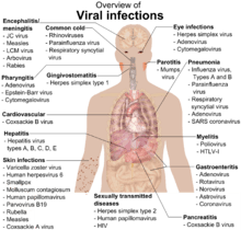
Examples of common human diseases caused by viruses include the common cold, influenza, chickenpox, and cold sores. Many serious diseases such as Ebola virus disease, AIDS, avian influenza, and SARS are caused by viruses. The relative ability of viruses to cause disease is described in terms of virulence. Other diseases are under investigation to discover if they have a virus as the causative agent, such as the possible connection between human herpesvirus 6 (HHV6) and neurological diseases such as multiple sclerosis and chronic fatigue syndrome.[146] There is controversy over whether the bornavirus, previously thought to cause neurological diseases in horses, could be responsible for psychiatric illnesses in humans.[147]
Viruses have different mechanisms by which they produce disease in an organism, which depends largely on the viral species. Mechanisms at the cellular level primarily include cell lysis, the breaking open and subsequent death of the cell. In multicellular organisms, if enough cells die, the whole organism will start to suffer the effects. Although viruses cause disruption of healthy homeostasis, resulting in disease, they may exist relatively harmlessly within an organism. An example would include the ability of the herpes simplex virus, which causes cold sores, to remain in a dormant state within the human body. This is called latency[148] and is a characteristic of the herpes viruses, including Epstein–Barr virus, which causes glandular fever, and varicella zoster virus, which causes chickenpox and shingles. Most people have been infected with at least one of these types of herpes virus.[149] However, these latent viruses might sometimes be beneficial, as the presence of the virus can increase immunity against bacterial pathogens, such as Yersinia pestis.[150]
Some viruses can cause lifelong or chronic infections, where the viruses continue to replicate in the body despite the host's defence mechanisms.[151] This is common in hepatitis B virus and hepatitis C virus infections. People chronically infected are known as carriers, as they serve as reservoirs of infectious virus.[152] In populations with a high proportion of carriers, the disease is said to be endemic.[153]
Epidemiology
Viral epidemiology is the branch of medical science that deals with the transmission and control of virus infections in humans. Transmission of viruses can be vertical, which means from mother to child, or horizontal, which means from person to person. Examples of vertical transmission include hepatitis B virus and HIV, where the baby is born already infected with the virus.[154] Another, more rare, example is the varicella zoster virus, which, although causing relatively mild infections in humans, can be fatal to the foetus and newborn baby.[155]
Horizontal transmission is the most common mechanism of spread of viruses in populations. Transmission can occur when: body fluids are exchanged during sexual activity, e.g., HIV; blood is exchanged by contaminated transfusion or needle sharing, e.g., hepatitis C; exchange of saliva by mouth, e.g., Epstein–Barr virus; contaminated food or water is ingested, e.g., norovirus; aerosols containing virions are inhaled, e.g., influenza virus; and insect vectors such as mosquitoes penetrate the skin of a host, e.g., dengue. The rate or speed of transmission of viral infections depends on factors that include population density, the number of susceptible individuals, (i.e., those not immune),[156] the quality of healthcare and the weather.[157]
Epidemiology is used to break the chain of infection in populations during outbreaks of viral diseases.[158] Control measures are used that are based on knowledge of how the virus is transmitted. It is important to find the source, or sources, of the outbreak and to identify the virus. Once the virus has been identified, the chain of transmission can sometimes be broken by vaccines. When vaccines are not available, sanitation and disinfection can be effective. Often, infected people are isolated from the rest of the community, and those that have been exposed to the virus are placed in quarantine.[159] To control the outbreak of foot-and-mouth disease in cattle in Britain in 2001, thousands of cattle were slaughtered.[160] Most viral infections of humans and other animals have incubation periods during which the infection causes no signs or symptoms.[161] Incubation periods for viral diseases range from a few days to weeks, but are known for most infections.[162] Somewhat overlapping, but mainly following the incubation period, there is a period of communicability — a time when an infected individual or animal is contagious and can infect another person or animal.[162] This, too, is known for many viral infections, and knowledge of the length of both periods is important in the control of outbreaks.[163] When outbreaks cause an unusually high proportion of cases in a population, community, or region, they are called epidemics. If outbreaks spread worldwide, they are called pandemics.[164]
Epidemics and pandemics
Native American populations were devastated by contagious diseases, in particular, smallpox, brought to the Americas by European colonists. It is unclear how many Native Americans were killed by foreign diseases after the arrival of Columbus in the Americas, but the numbers have been estimated to be close to 70% of the indigenous population. The damage done by this disease significantly aided European attempts to displace and conquer the native population.[165]
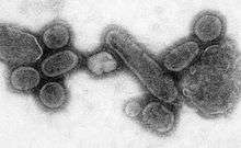
A pandemic is a worldwide epidemic. The 1918 flu pandemic, which lasted until 1919, was a category 5 influenza pandemic caused by an unusually severe and deadly influenza A virus. The victims were often healthy young adults, in contrast to most influenza outbreaks, which predominantly affect juvenile, elderly, or otherwise-weakened patients.[166] Older estimates say it killed 40–50 million people,[167] while more recent research suggests that it may have killed as many as 100 million people, or 5% of the world's population in 1918.[168]
Most researchers believe that HIV originated in sub-Saharan Africa during the 20th century;[169] it is now a pandemic, with an estimated 38.6 million people now living with the disease worldwide.[170] The Joint United Nations Programme on HIV/AIDS (UNAIDS) and the World Health Organization (WHO) estimate that AIDS has killed more than 25 million people since it was first recognised on 5 June 1981, making it one of the most destructive epidemics in recorded history.[171] In 2007 there were 2.7 million new HIV infections and 2 million HIV-related deaths.[172]
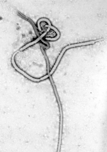
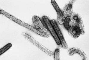
Several highly lethal viral pathogens are members of the Filoviridae. Filoviruses are filament-like viruses that cause viral hemorrhagic fever, and include ebolaviruses and marburgviruses. Marburg virus, first discovered in 1967, attracted widespread press attention in April 2005 for an outbreak in Angola.[173] Ebola Virus Disease has also caused intermittent outbreaks with high mortality rates since 1976 when it was first identified. The worst and most recent one is the West Africa epidemic.[174]
Cancer
Viruses are an established cause of cancer in humans and other species. Viral cancers occur only in a minority of infected persons (or animals). Cancer viruses come from a range of virus families, including both RNA and DNA viruses, and so there is no single type of "oncovirus" (an obsolete term originally used for acutely transforming retroviruses). The development of cancer is determined by a variety of factors such as host immunity[175] and mutations in the host.[176] Viruses accepted to cause human cancers include some genotypes of human papillomavirus, hepatitis B virus, hepatitis C virus, Epstein–Barr virus, Kaposi's sarcoma-associated herpesvirus and human T-lymphotropic virus. The most recently discovered human cancer virus is a polyomavirus (Merkel cell polyomavirus) that causes most cases of a rare form of skin cancer called Merkel cell carcinoma.[177] Hepatitis viruses can develop into a chronic viral infection that leads to liver cancer.[178][179] Infection by human T-lymphotropic virus can lead to tropical spastic paraparesis and adult T-cell leukaemia.[180] Human papillomaviruses are an established cause of cancers of cervix, skin, anus, and penis.[181] Within the Herpesviridae, Kaposi's sarcoma-associated herpesvirus causes Kaposi's sarcoma and body-cavity lymphoma, and Epstein–Barr virus causes Burkitt's lymphoma, Hodgkin's lymphoma, B lymphoproliferative disorder, and nasopharyngeal carcinoma.[182] Merkel cell polyomavirus closely related to SV40 and mouse polyomaviruses that have been used as animal models for cancer viruses for over 50 years.[183]
Host defence mechanisms
The body's first line of defence against viruses is the innate immune system. This comprises cells and other mechanisms that defend the host from infection in a non-specific manner. This means that the cells of the innate system recognise, and respond to, pathogens in a generic way, but, unlike the adaptive immune system, it does not confer long-lasting or protective immunity to the host.[184]
RNA interference is an important innate defence against viruses.[185] Many viruses have a replication strategy that involves double-stranded RNA (dsRNA). When such a virus infects a cell, it releases its RNA molecule or molecules, which immediately bind to a protein complex called a dicer that cuts the RNA into smaller pieces. A biochemical pathway – the RISC complex, is activated, which ensures cell survival by degrading the viral mRNA. Rotaviruses have evolved to avoid this defence mechanism by not uncoating fully inside the cell, and releasing newly produced mRNA through pores in the particle's inner capsid. Their genomic dsRNA remains protected inside the core of the virion.[186][187]
When the adaptive immune system of a vertebrate encounters a virus, it produces specific antibodies that bind to the virus and often render it non-infectious. This is called humoral immunity. Two types of antibodies are important. The first, called IgM, is highly effective at neutralising viruses but is produced by the cells of the immune system only for a few weeks. The second, called IgG, is produced indefinitely. The presence of IgM in the blood of the host is used to test for acute infection, whereas IgG indicates an infection sometime in the past.[188] IgG antibody is measured when tests for immunity are carried out.[189]
Antibodies can continue to be an effective defence mechanism even after viruses have managed to gain entry to the host cell. A protein that is in cells, called TRIM21, can attach to the antibodies on the surface of the virus particle. This primes the subsequent destruction of the virus by the enzymes of the cell's proteosome system.[190]
A second defence of vertebrates against viruses is called cell-mediated immunity and involves immune cells known as T cells. The body's cells constantly display short fragments of their proteins on the cell's surface, and, if a T cell recognises a suspicious viral fragment there, the host cell is destroyed by killer T cells and the virus-specific T-cells proliferate. Cells such as the macrophage are specialists at this antigen presentation.[191] The production of interferon is an important host defence mechanism. This is a hormone produced by the body when viruses are present. Its role in immunity is complex; it eventually stops the viruses from reproducing by killing the infected cell and its close neighbours.[192]
Not all virus infections produce a protective immune response in this way. HIV evades the immune system by constantly changing the amino acid sequence of the proteins on the surface of the virion. This is known as "escape mutation" as the viral epitopes escape recognition by the host immune response. These persistent viruses evade immune control by sequestration, blockade of antigen presentation, cytokine resistance, evasion of natural killer cell activities, escape from apoptosis, and antigenic shift.[193] Other viruses, called neurotropic viruses, are disseminated by neural spread where the immune system may be unable to reach them.
Prevention and treatment
Because viruses use vital metabolic pathways within host cells to replicate, they are difficult to eliminate without using drugs that cause toxic effects to host cells in general. The most effective medical approaches to viral diseases are vaccinations to provide immunity to infection, and antiviral drugs that selectively interfere with viral replication.
Vaccines
Vaccination is a cheap and effective way of preventing infections by viruses. Vaccines were used to prevent viral infections long before the discovery of the actual viruses. Their use has resulted in a dramatic decline in morbidity (illness) and mortality (death) associated with viral infections such as polio, measles, mumps and rubella.[194] Smallpox infections have been eradicated.[195] Vaccines are available to prevent over thirteen viral infections of humans,[196] and more are used to prevent viral infections of animals.[197] Vaccines can consist of live-attenuated or killed viruses, or viral proteins (antigens).[198] Live vaccines contain weakened forms of the virus, which do not cause the disease but, nonetheless, confer immunity. Such viruses are called attenuated. Live vaccines can be dangerous when given to people with a weak immunity (who are described as immunocompromised), because in these people, the weakened virus can cause the original disease.[199] Biotechnology and genetic engineering techniques are used to produce subunit vaccines. These vaccines use only the capsid proteins of the virus. Hepatitis B vaccine is an example of this type of vaccine.[200] Subunit vaccines are safe for immunocompromised patients because they cannot cause the disease.[201] The yellow fever virus vaccine, a live-attenuated strain called 17D, is probably the safest and most effective vaccine ever generated.[202]
Antiviral drugs

Antiviral drugs are often nucleoside analogues (fake DNA building-blocks), which viruses mistakenly incorporate into their genomes during replication. The life-cycle of the virus is then halted because the newly synthesised DNA is inactive. This is because these analogues lack the hydroxyl groups, which, along with phosphorus atoms, link together to form the strong "backbone" of the DNA molecule. This is called DNA chain termination.[203] Examples of nucleoside analogues are aciclovir for Herpes simplex virus infections and lamivudine for HIV and Hepatitis B virus infections. Aciclovir is one of the oldest and most frequently prescribed antiviral drugs.[204] Other antiviral drugs in use target different stages of the viral life cycle. HIV is dependent on a proteolytic enzyme called the HIV-1 protease for it to become fully infectious. There is a large class of drugs called protease inhibitors that inactivate this enzyme.
Hepatitis C is caused by an RNA virus. In 80% of people infected, the disease is chronic, and without treatment, they are infected for the remainder of their lives. However, there is now an effective treatment that uses the nucleoside analogue drug ribavirin combined with interferon.[205] The treatment of chronic carriers of the hepatitis B virus by using a similar strategy using lamivudine has been developed.[206]
Infection in other species
Viruses infect all cellular life and, although viruses occur universally, each cellular species has its own specific range that often infect only that species.[207] Some viruses, called satellites, can replicate only within cells that have already been infected by another virus.[58]
Animal viruses
Viruses are important pathogens of livestock. Diseases such as foot-and-mouth disease and bluetongue are caused by viruses.[208] Companion animals such as cats, dogs, and horses, if not vaccinated, are susceptible to serious viral infections. Canine parvovirus is caused by a small DNA virus and infections are often fatal in pups.[209] Like all invertebrates, the honey bee is susceptible to many viral infections.[210] However, most viruses co-exist harmlessly in their host and cause no signs or symptoms of disease.[2]
Plant viruses

There are many types of plant virus, but often they cause only a loss of yield, and it is not economically viable to try to control them. Plant viruses are often spread from plant to plant by organisms, known as vectors. These are normally insects, but some fungi, nematode worms, and single-celled organisms have been shown to be vectors. When control of plant virus infections is considered economical, for perennial fruits, for example, efforts are concentrated on killing the vectors and removing alternate hosts such as weeds.[211] Plant viruses cannot infect humans and other animals because they can reproduce only in living plant cells.[212]
Plants have elaborate and effective defence mechanisms against viruses. One of the most effective is the presence of so-called resistance (R) genes. Each R gene confers resistance to a particular virus by triggering localised areas of cell death around the infected cell, which can often be seen with the unaided eye as large spots. This stops the infection from spreading.[213] RNA interference is also an effective defence in plants.[214] When they are infected, plants often produce natural disinfectants that kill viruses, such as salicylic acid, nitric oxide, and reactive oxygen molecules.[215]
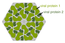
Plant virus particles or virus-like particles (VLPs) have applications in both biotechnology and nanotechnology. The capsids of most plant viruses are simple and robust structures and can be produced in large quantities either by the infection of plants or by expression in a variety of heterologous systems. Plant virus particles can be modified genetically and chemically to encapsulate foreign material and can be incorporated into supramolecular structures for use in biotechnology.[216]
Bacterial viruses
Bacteriophages are a common and diverse group of viruses and are the most abundant form of biological entity in aquatic environments – there are up to ten times more of these viruses in the oceans than there are bacteria,[217] reaching levels of 250,000,000 bacteriophages per millilitre of seawater.[218] These viruses infect specific bacteria by binding to surface receptor molecules and then entering the cell. Within a short amount of time, in some cases just minutes, bacterial polymerase starts translating viral mRNA into protein. These proteins go on to become either new virions within the cell, helper proteins, which help assembly of new virions, or proteins involved in cell lysis. Viral enzymes aid in the breakdown of the cell membrane, and, in the case of the T4 phage, in just over twenty minutes after injection over three hundred phages could be released.[219]
The major way bacteria defend themselves from bacteriophages is by producing enzymes that destroy foreign DNA. These enzymes, called restriction endonucleases, cut up the viral DNA that bacteriophages inject into bacterial cells.[220] Bacteria also contain a system that uses CRISPR sequences to retain fragments of the genomes of viruses that the bacteria have come into contact with in the past, which allows them to block the virus's replication through a form of RNA interference.[221][222] This genetic system provides bacteria with acquired immunity to infection.[223]
Archaean viruses
Some viruses replicate within archaea: these are double-stranded DNA viruses with unusual and sometimes unique shapes.[5][89] These viruses have been studied in most detail in the thermophilic archaea, particularly the orders Sulfolobales and Thermoproteales.[224] Defences against these viruses involve RNA interference from repetitive DNA sequences within archaean genomes that are related to the genes of the viruses.[225][226] Most archaea have CRISPR–Cas systems as an adaptive defence against viruses. These enable archaea to retain sections of viral DNA, which are then used to target and eliminate subsequent infections by the virus using a process similar to RNA interference.[227]
Role in aquatic ecosystems
A teaspoon of seawater contains about one million viruses.[228] Most of these are bacteriophages, which are harmless to plants and animals, and are in fact essential to the regulation of saltwater and freshwater ecosystems.[229] They infect and destroy bacteria in aquatic microbial communities, and are the most important mechanism of recycling carbon in the marine environment. The organic molecules released from the dead bacterial cells stimulate fresh bacterial and algal growth.[230] Viral activity may also contribute to the biological pump, the process whereby carbon is sequestered in the deep ocean.[231]
Microorganisms constitute more than 90% of the biomass in the sea. It is estimated that viruses kill approximately 20% of this biomass each day and that there are 15 times as many viruses in the oceans as there are bacteria and archaea. Viruses are the main agents responsible for the rapid destruction of harmful algal blooms,[232] which often kill other marine life.[233] The number of viruses in the oceans decreases further offshore and deeper into the water, where there are fewer host organisms.[231]
Like any organism, marine mammals are susceptible to viral infections. In 1988 and 2002, thousands of harbour seals were killed in Europe by phocine distemper virus.[234] Many other viruses, including caliciviruses, herpesviruses, adenoviruses and parvoviruses, circulate in marine mammal populations.[231]
Role in evolution
Viruses are an important natural means of transferring genes between different species, which increases genetic diversity and drives evolution.[7] It is thought that viruses played a central role in the early evolution, before the diversification of bacteria, archaea and eukaryotes, at the time of the last universal common ancestor of life on Earth.[235] Viruses are still one of the largest reservoirs of unexplored genetic diversity on Earth.[231]
Applications
Life sciences and medicine

Viruses are important to the study of molecular and cell biology as they provide simple systems that can be used to manipulate and investigate the functions of cells.[236] The study and use of viruses have provided valuable information about aspects of cell biology.[237] For example, viruses have been useful in the study of genetics and helped our understanding of the basic mechanisms of molecular genetics, such as DNA replication, transcription, RNA processing, translation, protein transport, and immunology.
Geneticists often use viruses as vectors to introduce genes into cells that they are studying. This is useful for making the cell produce a foreign substance, or to study the effect of introducing a new gene into the genome. In similar fashion, virotherapy uses viruses as vectors to treat various diseases, as they can specifically target cells and DNA. It shows promising use in the treatment of cancer and in gene therapy. Eastern European scientists have used phage therapy as an alternative to antibiotics for some time, and interest in this approach is increasing, because of the high level of antibiotic resistance now found in some pathogenic bacteria.[238] Expression of heterologous proteins by viruses is the basis of several manufacturing processes that are currently being used for the production of various proteins such as vaccine antigens and antibodies. Industrial processes have been recently developed using viral vectors and a number of pharmaceutical proteins are currently in pre-clinical and clinical trials.[239]
Virotherapy
Virotherapy involves the use of genetically modified viruses to treat diseases.[240] Viruses have been modified by scientists to reproduce in cancer cells and destroy them but not infect healthy cells. Talimogene laherparepvec (T-VEC), for example, is a modified herpes simplex virus that has had a gene, which is required for viruses to replicate in healthy cells, deleted and replaced with a human gene (GM-CSF) that stimulates immunity. When this virus infects cancer cells, it destroys them and in doing so the presence the GM-CSF gene attracts dendritic cells from the surrounding tissues of the body. The dendritic cells process the dead cancer cells and present components of them to other cells of the immune system.[241] Having completed successful clinical trials, this virus is expected to gain approval for the treatment of a skin cancer called melanoma in late 2015.[242] Viruses that have been reprogrammed to kill cancer cells are called oncolytic viruses.[243]
Materials science and nanotechnology
Current trends in nanotechnology promise to make much more versatile use of viruses. From the viewpoint of a materials scientist, viruses can be regarded as organic nanoparticles. Their surface carries specific tools designed to cross the barriers of their host cells. The size and shape of viruses, and the number and nature of the functional groups on their surface, is precisely defined. As such, viruses are commonly used in materials science as scaffolds for covalently linked surface modifications. A particular quality of viruses is that they can be tailored by directed evolution. The powerful techniques developed by life sciences are becoming the basis of engineering approaches towards nanomaterials, opening a wide range of applications far beyond biology and medicine.[244]
Because of their size, shape, and well-defined chemical structures, viruses have been used as templates for organising materials on the nanoscale. Recent examples include work at the Naval Research Laboratory in Washington, D.C., using Cowpea mosaic virus (CPMV) particles to amplify signals in DNA microarray based sensors. In this application, the virus particles separate the fluorescent dyes used for signalling to prevent the formation of non-fluorescent dimers that act as quenchers.[245] Another example is the use of CPMV as a nanoscale breadboard for molecular electronics.[246]
Synthetic viruses
Many viruses can be synthesised de novo ("from scratch") and the first synthetic virus was created in 2002.[247] Although somewhat of a misconception, it is not the actual virus that is synthesised, but rather its DNA genome (in case of a DNA virus), or a cDNA copy of its genome (in case of RNA viruses). For many virus families the naked synthetic DNA or RNA (once enzymatically converted back from the synthetic cDNA) is infectious when introduced into a cell. That is, they contain all the necessary information to produce new viruses. This technology is now being used to investigate novel vaccine strategies.[248] The ability to synthesise viruses has far-reaching consequences, since viruses can no longer be regarded as extinct, as long as the information of their genome sequence is known and permissive cells are available. As of March 2014, the full-length genome sequences of 3843 different viruses, including smallpox, are publicly available in an online database maintained by the National Institutes of Health.[249]
Weapons
The ability of viruses to cause devastating epidemics in human societies has led to the concern that viruses could be weaponised for biological warfare. Further concern was raised by the successful recreation of the infamous 1918 influenza virus in a laboratory.[250] Smallpox virus devastated numerous societies throughout history before its eradication. There are only two centres in the world that are authorised by the WHO to keep stocks of smallpox virus: the Vector Institute in Russia and the Centers for Disease Control and Prevention in the United States.[251] Fears that it may be used as a weapon may not be totally unfounded.[251] As the vaccine for smallpox sometimes had severe side-effects, it is no longer used routinely in any country. Thus, much of the modern human population has almost no established resistance to smallpox, and would be vulnerable to the virus.[251]
See also
- Cross-species transmission
- Non-cellular life
- Human timeline
- Nature timeline
- Viral metagenomics
- Viroplasm
- Zoonosis
References
Notes
- 1 2 Koonin EV, Senkevich TG, Dolja VV. The ancient Virus World and evolution of cells. Biol. Direct. 2006;1:29. doi:10.1186/1745-6150-1-29. PMID 16984643.
- 1 2 3 Dimmock p. 4
- 1 2 3 Dimmock p. 49
- 1 2 Breitbart M, Rohwer F. Here a virus, there a virus, everywhere the same virus?. Trends Microbiol. 2005;13(6):278–84. doi:10.1016/j.tim.2005.04.003. PMID 15936660.
- 1 2 Lawrence CM, Menon S, Eilers BJ, et al.. Structural and functional studies of archaeal viruses. J. Biol. Chem.. 2009;284(19):12599–603. doi:10.1074/jbc.R800078200. PMID 19158076.
- ↑ Edwards RA, Rohwer F. Viral metagenomics. Nature Reviews Microbiology. 2005;3(6):504–10. doi:10.1038/nrmicro1163. PMID 15886693.
- 1 2 Canchaya C, Fournous G, Chibani-Chennoufi S, Dillmann ML, Brüssow H. Phage as agents of lateral gene transfer. Current Opinion in Microbiology. 2003;6(4):417–24. doi:10.1016/S1369-5274(03)00086-9. PMID 12941415.
- 1 2 Rybicki, EP. The classification of organisms at the edge of life, or problems with virus systematics. S Afr J Sci. 1990;86:182–186.
- ↑ Koonin, E. V.; Starokadomskyy, P. (7 March 2016). "Are viruses alive? The replicator paradigm sheds decisive light on an old but misguided question.". Stud Hist Philos Biol Biomed Sci. doi:10.1016/j.shpsc.2016.02.016. PMID 26965225.
- ↑ Shors pp. 49–50
- ↑ "virus, n." OED Online. Oxford University Press, March 2015. Web. 23 March 2015.
- 1 2 Harper D. The Online Etymology Dictionary. virus; 2011 [Retrieved 2014-12-19].
- ↑ "virulent, adj." OED Online. Oxford University Press, March 2015. Web. 23 March 2015.
- ↑ Harper D. The Online Etymology Dictionary. virulent; 2011 [Retrieved 2014-12-19].
- ↑ e.g. Michael Worboys: Cambridge History of Medicine: Spreading Germs: Disease Theories and Medical Practice in Britain, 1865-1900, Cambridge University Press, 2000, p. 204
- ↑ e.g. Karsten Buschard & Rikke Thon: Diabetic Animal Models. In: Handbook of Laboratory Animal Science. Second Edition. Volume II: Animal Models, edited by Jann Hau & Gerald L. Van Hoosier Jr., CRC Press, 2003, p. 163 & 166
- ↑ William T. Stearn: Botanical Latin. History, Grammar, Syntax, Terminology and Vocabulary. David & Charles, third edition, 1983. Quote: "Virus: virus (s.n. II), gen. sing. viri, nom. pl. vira, gen. pl. vīrorum (to be distinguished from virorum, of men)."
- ↑ Pons: virus
- ↑ Harper D. The Online Etymology Dictionary. viral; 2011 [Retrieved 2014-12-19].
- ↑ Harper D. The Online Etymology Dictionary. virion; 2011 [Retrieved 2014-12-19].
- ↑ Casjens S. In: Mahy BWJ and Van Regenmortel MHV. Desk Encyclopedia of General Virology. Boston: Academic Press; 2010. ISBN 0-12-375146-2. p. 167.
- ↑ Bordenave G. Louis Pasteur (1822–1895). Microbes and Infection / Institut Pasteur. 2003;5(6):553–60. doi:10.1016/S1286-4579(03)00075-3. PMID 12758285.
- ↑ Shors pp. 76–77
- 1 2 3 Collier p. 3
- ↑ Dimmock p.4–5
- ↑ Fenner F.. In: Mahy B. W. J. and Van Regenmortal M. H. V.. Desk Encyclopedia of General Virology. 1 ed. Oxford, UK: Academic Press; 2009. ISBN 0-12-375146-2. p. 15.
- ↑ Shors p. 589
- ↑ D'Herelle F. On an invisible microbe antagonistic toward dysenteric bacilli: brief note by Mr. F. D'Herelle, presented by Mr. Roux. Research in Microbiology. 2007;158(7):553–4. doi:10.1016/j.resmic.2007.07.005. PMID 17855060.
- ↑ Steinhardt E, Israeli C, Lambert R.A.. Studies on the cultivation of the virus of vaccinia. J. Inf Dis.. 1913;13(2):294–300. doi:10.1093/infdis/13.2.294.
- ↑ Collier p. 4
- ↑ Goodpasture EW, Woodruff AM, Buddingh GJ. The cultivation of vaccine and other viruses in the chorioallantoic membrane of chick embryos. Science. 1931;74(1919):371–372. doi:10.1126/science.74.1919.371. PMID 17810781. Bibcode:1931Sci....74..371G.
- ↑ Rosen, FS. Isolation of poliovirus—John Enders and the Nobel Prize. New England Journal of Medicine. 2004;351(15):1481–83. doi:10.1056/NEJMp048202. PMID 15470207.
- ↑ From Nobel Lectures, Physics 1981–1990, (1993) Editor-in-Charge Tore Frängsmyr, Editor Gösta Ekspång, World Scientific Publishing Co., Singapore.
- In 1887, Buist visualised one of the largest, Vaccinia virus, by optical microscopy after staining it. Vaccinia was not known to be a virus at that time. (Buist J.B. Vaccinia and Variola: a study of their life history Churchill, London)
- ↑ Stanley WM, Loring HS. The isolation of crystalline tobacco mosaic virus protein from diseased tomato plants. Science. 1936;83(2143):85. doi:10.1126/science.83.2143.85. PMID 17756690. Bibcode:1936Sci....83...85S.
- ↑ Stanley WM, Lauffer MA. Disintegration of tobacco mosaic virus in urea solutions. Science. 1939;89(2311):345–347. doi:10.1126/science.89.2311.345. PMID 17788438. Bibcode:1939Sci....89..345S.
- ↑ Creager AN, Morgan GJ. After the double helix: Rosalind Franklin's research on Tobacco mosaic virus. Isis. 2008;99(2):239–72. doi:10.1086/588626. PMID 18702397.
- ↑ Dimmock p. 12
- ↑ Norrby E. Nobel Prizes and the emerging virus concept. Arch. Virol.. 2008;153(6):1109–23. doi:10.1007/s00705-008-0088-8. PMID 18446425.
- ↑ Collier p. 745
- 1 2 Temin HM, Baltimore D. RNA-directed DNA synthesis and RNA tumor viruses. Adv. Virus Res.. 1972 [Retrieved 16 September 2008];17:129–86. doi:10.1016/S0065-3527(08)60749-6. PMID 4348509.
- ↑ Barré-Sinoussi, F. et al.. Isolation of a T-lymphotropic retrovirus from a patient at risk for acquired immune deficiency syndrome (AIDS). Science. 1983;220(4599):868–871. doi:10.1126/science.6189183. PMID 6189183. Bibcode:1983Sci...220..868B.
- ↑ Choo QL, Kuo G, Weiner AJ, Overby LR, Bradley DW, Houghton M (April 1989). "Isolation of a cDNA clone derived from a blood-borne non-A, non-B viral hepatitis genome". Science. 244 (4902): 359–62. doi:10.1126/science.2523562. PMID 2523562.
- ↑ Houghton M (November 2009). "The long and winding road leading to the identification of the hepatitis C virus". Journal of Hepatology. 51 (5): 939–48. doi:10.1016/j.jhep.2009.08.004. PMID 19781804.
- ↑ Iyer LM, Balaji S, Koonin EV, Aravind L. Evolutionary genomics of nucleo-cytoplasmic large DNA viruses. Virus Res.. 2006;117(1):156–84. doi:10.1016/j.virusres.2006.01.009. PMID 16494962.
- 1 2 Sanjuán R, Nebot MR, Chirico N, Mansky LM, Belshaw R. Viral mutation rates. Journal of Virology. 2010;84(19):9733–48. doi:10.1128/JVI.00694-10. PMID 20660197.
- ↑ Shors pp. 14–16
- ↑ Collier pp. 11–21
- 1 2 Dimmock p. 16
- ↑ Collier p. 11
- 1 2 3 4 Mahy WJ & Van Regenmortel MHV (eds). Desk Encyclopedia of General Virology. Oxford: Academic Press; 2009. ISBN 0-12-375146-2. p. 24.
- ↑ Shors p. 574
- ↑ The origin and behavior of mutable loci in maize. Proc Natl Acad Sci U S A.. 1950;36(6):344–55. doi:10.1073/pnas.36.6.344. PMID 15430309. Bibcode:1950PNAS...36..344M.
- ↑ Collier pp. 11–12
- ↑ Dimmock p. 55
- ↑ Shors 551–3
- ↑ Tsagris EM, de Alba AE, Gozmanova M, Kalantidis K. Viroids. Cell. Microbiol.. 2008;10(11):2168. doi:10.1111/j.1462-5822.2008.01231.x. PMID 18764915.
- ↑ Shors p. 492–3
- 1 2 La Scola B, Desnues C, Pagnier I, Robert C, Barrassi L, Fournous G, Merchat M, Suzan-Monti M, Forterre P, Koonin E, Raoult D. The virophage as a unique parasite of the giant mimivirus. Nature. 2008;455(7209):100–4. doi:10.1038/nature07218. PMID 18690211. Bibcode:2008Natur.455..100L.
- ↑ Collier p. 777
- ↑ Dimmock p. 55–7
- 1 2 Mahy WJ & Van Regenmortel MHV (eds). Desk Encyclopedia of General Virology. Oxford: Academic Press; 2009. ISBN 0-12-375146-2. p. 28.
- 1 2 Mahy WJ & Van Regenmortel MHV (eds). Desk Encyclopedia of General Virology. Oxford: Academic Press; 2009. ISBN 0-12-375146-2. p. 26.
- ↑ Dimmock pp. 15–16
- ↑ Liberski PP. Prion diseases: a riddle wrapped in a mystery inside an enigma. Folia Neuropathol. 2008;46(2):93–116. PMID 18587704.
- ↑ Belay ED and Schonberger LB. Desk Encyclopedia of Human and Medical Virology. Boston: Academic Press; 2009. ISBN 0-12-375147-0. p. 497–504.
- ↑ Lupi O, Dadalti P, Cruz E, Goodheart C. Did the first virus self-assemble from self-replicating prion proteins and RNA?. Med. Hypotheses. 2007;69(4):724–30. doi:10.1016/j.mehy.2007.03.031. PMID 17512677.
- ↑ Are viruses alive? The replicator paradigm sheds decisive light on an old but misguided question. Studies in History and Philosophy of Biological and Biomedical Sciences. 2016. doi:10.1016/j.shpsc.2016.02.016. PMID 26965225.
- ↑ Holmes EC. Viral evolution in the genomic age. PLoS Biol.. 2007;5(10):e278. doi:10.1371/journal.pbio.0050278. PMID 17914905.
- ↑ Wimmer E, Mueller S, Tumpey TM, Taubenberger JK. Synthetic viruses: a new opportunity to understand and prevent viral disease. Nature Biotechnology. 2009;27(12):1163–72. doi:10.1038/nbt.1593. PMID 20010599.
- ↑ Horn M. Chlamydiae as symbionts in eukaryotes. Annual Review of Microbiology. 2008;62:113–31. doi:10.1146/annurev.micro.62.081307.162818. PMID 18473699.
- ↑ Ammerman NC, Beier-Sexton M, Azad AF. Laboratory maintenance of Rickettsia rickettsii. Current Protocols in Microbiology. 2008;Chapter 3:Unit 3A.5. doi:10.1002/9780471729259.mc03a05s11. PMID 19016440.
- 1 2 Collier pp. 33–55
- ↑ Collier pp. 33–37
- ↑ Kiselev NA, Sherman MB, Tsuprun VL. Negative staining of proteins. Electron Microsc. Rev.. 1990;3(1):43–72. doi:10.1016/0892-0354(90)90013-I. PMID 1715774.
- ↑ Collier p. 40
- ↑ Caspar DL, Klug A. Physical principles in the construction of regular viruses. Cold Spring Harb. Symp. Quant. Biol.. 1962;27:1–24. doi:10.1101/sqb.1962.027.001.005. PMID 14019094.
- ↑ Crick FH, Watson JD. Structure of small viruses. Nature. 1956;177(4506):473–5. doi:10.1038/177473a0. PMID 13309339. Bibcode:1956Natur.177..473C.
- ↑ Manipulation of individual viruses: friction and mechanical properties. Biophysical Journal. 1997;72(3):1396–1403. doi:10.1016/S0006-3495(97)78786-1. PMID 9138585. Bibcode:1997BpJ....72.1396F.
- ↑ Imaging of viruses by atomic force microscopy. J Gen Virol. 2001;82(9):2025–2034. PMID 11514711.
- ↑ Collier p. 37
- ↑ Collier pp. 40, 42
- ↑ Casens, S. Desk Encyclopedia of General Virology. Boston: Academic Press; 2009. ISBN 0-12-375146-2. p. 167–174.
- ↑ Collier pp. 42–43
- ↑ Rossmann MG, Mesyanzhinov VV, Arisaka F, Leiman PG. The bacteriophage T4 DNA injection machine. Current Opinion in Structural Biology. 2004;14(2):171–80. doi:10.1016/j.sbi.2004.02.001. PMID 15093831.
- ↑ Long GW, Nobel J, Murphy FA, Herrmann KL, Lourie B. Experience with electron microscopy in the differential diagnosis of smallpox. Appl Microbiol. 1970;20(3):497–504. PMID 4322005.
- ↑ Suzan-Monti M, La Scola B, Raoult D. Genomic and evolutionary aspects of Mimivirus. Virus Research. 2006;117(1):145–155. doi:10.1016/j.virusres.2005.07.011. PMID 16181700.
- ↑ Arslan D, Legendre M, Seltzer V, Abergel C, Claverie JM. Distant Mimivirus relative with a larger genome highlights the fundamental features of Megaviridae. Proceedings of the National Academy of Sciences of the United States of America. 2011;108(42):17486–91. doi:10.1073/pnas.1110889108. PMID 21987820. Bibcode:2011PNAS..10817486A.
- ↑ Philippe Nadège; et al. (2013). "Pandoraviruses: amoeba viruses with genomes up to 2.5 Mb reaching that of parasitic eukaryotes". Science. 341 (6143): 281–286. Bibcode:2013Sci...341..281P. doi:10.1126/science.1239181. PMID 23869018.
- 1 2 Prangishvili D, Forterre P, Garrett RA. Viruses of the Archaea: a unifying view. Nature Reviews Microbiology. 2006;4(11):837–48. doi:10.1038/nrmicro1527. PMID 17041631.
- ↑ NCBI Viral Genome database
- ↑ Pennisi E (2011). "Going Viral: Exploring the Role Of Viruses in Our Bodies". Science. 331 (6024): 1513. Bibcode:2011Sci...331.1513P. doi:10.1126/science.331.6024.1513. PMID 21436418.
- ↑ Shi, Mang; Lin, Xian-Dan; Tian, Jun-Hua; Chen, Liang-Jun; Chen, Xiao; Li, Ci-Xiu; Qin, Xin-Cheng; Li, Jun; Cao, Jian-Ping (2016). "Redefining the invertebrate RNA virosphere". Nature. doi:10.1038/nature20167.
- 1 2 3 Collier pp. 96–99
- ↑ Saunders, Venetia A.; Carter, John. Virology: principles and applications. Chichester: John Wiley & Sons; 2007. ISBN 0-470-02387-2. p. 72.
- ↑ Belyi VA, Levine AJ, Skalka AM (2010). "Sequences from ancestral single-stranded DNA viruses in vertebrate genomes: the parvoviridae and circoviridae are more than 40 to 50 million years old". J. Virol. 84 (23): 12458–62. doi:10.1128/JVI.01789-10. PMC 2976387
 . PMID 20861255.
 - ↑ Philippe N, Legendre M, Doutre G, Couté Y, Poirot O, Lescot M, Arslan D, Seltzer V, Bertaux L, Bruley C, Garin J, Claverie JM, Abergel C (2013). "Pandoraviruses: amoeba viruses with genomes up to 2.5 Mb reaching that of parasitic eukaryotes". Science. 341 (6143): 281–6. Bibcode:2013Sci...341..281P. doi:10.1126/science.1239181. PMID 23869018.
- ↑ Pressing J, Reanney DC. Divided genomes and intrinsic noise. J Mol Evol. 1984;20(2):135–46. doi:10.1007/BF02257374. PMID 6433032.
- ↑ Duffy S, Holmes EC. Validation of high rates of nucleotide substitution in geminiviruses: phylogenetic evidence from East African cassava mosaic viruses. The Journal of General Virology. 2009;90(Pt 6):1539–47. doi:10.1099/vir.0.009266-0. PMID 19264617.
- ↑ Sandbulte MR, Westgeest KB, Gao J, Xu X, Klimov AI, Russell CA, Burke DF, Smith DJ, Fouchier RA, Eichelberger MC. Discordant antigenic drift of neuraminidase and hemagglutinin in H1N1 and H3N2 influenza viruses. Proceedings of the National Academy of Sciences of the United States of America. 2011;108(51):20748–53. doi:10.1073/pnas.1113801108. PMID 22143798. Bibcode:2011PNAS..10820748S.
- ↑ Moss RB, Davey RT, Steigbigel RT, Fang F. Targeting pandemic influenza: a primer on influenza antivirals and drug resistance. The Journal of Antimicrobial Chemotherapy. 2010;65(6):1086–93. doi:10.1093/jac/dkq100. PMID 20375034.
- ↑ Hampson AW, Mackenzie JS. The influenza viruses. Med. J. Aust.. 2006;185(10 Suppl):S39–43. PMID 17115950.
- ↑ Metzner KJ. Detection and significance of minority quasispecies of drug-resistant HIV-1. J HIV Ther. 2006;11(4):74–81. PMID 17578210.
- ↑ Goudsmit, Jaap. Viral Sex. Oxford Univ Press, 1998.ISBN 978-0-19-512496-5 ISBN 0-19-512496-0
- ↑ Worobey M, Holmes EC. Evolutionary aspects of recombination in RNA viruses. J. Gen. Virol.. 1999;80 ( Pt 10):2535–43. PMID 10573145.
- ↑ Lukashev AN. Role of recombination in evolution of enteroviruses. Rev. Med. Virol.. 2005;15(3):157–67. doi:10.1002/rmv.457. PMID 15578739.
- ↑ Umene K. Mechanism and application of genetic recombination in herpesviruses. Rev. Med. Virol.. 1999;9(3):171–82. doi:10.1002/(SICI)1099-1654(199907/09)9:3<171::AID-RMV243>3.0.CO;2-A. PMID 10479778.
- ↑ Collier pp. 75–91
- ↑ Dimmock p. 70
- ↑ Boevink P, Oparka KJ. Virus-host interactions during movement processes. Plant Physiol.. 2005;138(4):1815–21. doi:10.1104/pp.105.066761. PMID 16172094.
- ↑ Dimmock p. 71
- ↑ Barman S, Ali A, Hui EK, Adhikary L, Nayak DP. Transport of viral proteins to the apical membranes and interaction of matrix protein with glycoproteins in the assembly of influenza viruses. Virus Res.. 2001;77(1):61–9. doi:10.1016/S0168-1702(01)00266-0. PMID 11451488.
- ↑ Shors pp. 60, 597
- ↑ Dimmock, Chapter 15, Mechanisms in virus latentcy, pp.243–259
- ↑ Dimmock 185–187
- ↑ Shors p. 54; Collier p. 78
- ↑ Collier p. 79
- ↑ Staginnus C, Richert-Pöggeler KR. Endogenous pararetroviruses: two-faced travelers in the plant genome. Trends in Plant Science. 2006;11(10):485–91. doi:10.1016/j.tplants.2006.08.008. PMID 16949329.
- ↑ Collier pp. 88–89
- ↑ Collier pp. 115–146
- ↑ Collier p. 115
- ↑ Roulston A, Marcellus RC, Branton PE. Viruses and apoptosis. Annu. Rev. Microbiol.. 1999;53:577–628. doi:10.1146/annurev.micro.53.1.577. PMID 10547702.
- ↑ Alwine JC. Modulation of host cell stress responses by human cytomegalovirus. Curr. Top. Microbiol. Immunol.. 2008;325:263–79. doi:10.1007/978-3-540-77349-8_15. PMID 18637511.
- ↑ Sinclair J. Human cytomegalovirus: Latency and reactivation in the myeloid lineage. J. Clin. Virol.. 2008;41(3):180–5. doi:10.1016/j.jcv.2007.11.014. PMID 18164651.
- ↑ Jordan MC, Jordan GW, Stevens JG, Miller G. Latent herpesviruses of humans. Annals of Internal Medicine. 1984;100(6):866–80. doi:10.7326/0003-4819-100-6-866. PMID 6326635.
- ↑ Sissons JG, Bain M, Wills MR. Latency and reactivation of human cytomegalovirus. J. Infect.. 2002;44(2):73–7. doi:10.1053/jinf.2001.0948. PMID 12076064.
- ↑ Barozzi P, Potenza L, Riva G, Vallerini D, Quadrelli C, Bosco R, Forghieri F, Torelli G, Luppi M. B cells and herpesviruses: a model of lymphoproliferation. Autoimmun Rev. 2007;7(2):132–6. doi:10.1016/j.autrev.2007.02.018. PMID 18035323.
- ↑ Subramanya D, Grivas PD. HPV and cervical cancer: updates on an established relationship. Postgrad Med. 2008;120(4):7–13. doi:10.3810/pgm.2008.11.1928. PMID 19020360.
- ↑ Crawford, Dorothy H.. Viruses: A Very Short Introduction. Oxford University Press, USA; 2011. ISBN 0-19-957485-5. p. 16.
- ↑ Shors p. 388
- ↑ Shors p. 353
- ↑ Dimmock p. 272
- ↑ Baggesen DL, Sørensen G, Nielsen EM, Wegener HC. Phage typing of Salmonella Typhimurium – is it still a useful tool for surveillance and outbreak investigation?. Eurosurveillance. 2010 [Retrieved 2014-12-19];15(4):19471. PMID 20122382.
- ↑ Lwoff A, Horne RW, Tournier P. A virus system. C. R. Hebd. Seances Acad. Sci.. 1962;254:4225–7. French. PMID 14467544.
- ↑ Lwoff A, Horne R, Tournier P. A system of viruses. Cold Spring Harb. Symp. Quant. Biol.. 1962;27:51–5. doi:10.1101/sqb.1962.027.001.008. PMID 13931895.
- 1 2 King AMQ, Lefkowitz E, Adams MJ, Carstens EB. Virus Taxonomy: Ninth Report of the International Committee on Taxonomy of Viruses. Elsevier; 2011. ISBN 0-12-384684-6. p. 6.
- ↑ Adams, MJ.; Lefkowitz, EJ.; King, AM.; Carstens, EB. (Dec 2013). "Recently agreed changes to the International Code of Virus Classification and Nomenclature". Archives of Virology. 158 (12): 2633–9. doi:10.1007/s00705-013-1749-9. PMID 23836393.
- As defined therein, "A species is the lowest taxonomic level in the hierarchy approved by the ICTV. A species is a monophyletic group of viruses whose properties can be distinguished from those of other species by multiple criteria."
- ↑ Delwart EL. Viral metagenomics. Rev. Med. Virol.. 2007;17(2):115–31. doi:10.1002/rmv.532. PMID 17295196.
- ↑ Virus Taxonomy 2013. International Committee on Taxonomy of Viruses. Retrieved on 24 December 2011.
- ↑ ICTV Master Species List 2013 v2
- This Excel file contains the official ICTV Master Species list for 2014 and lists all approved virus taxa. This is version 11 of the MSL published on 24 August 2014. Retrieved on 24 December 2014
- ↑ Baltimore D. The strategy of RNA viruses. Harvey Lect.. 1974;70 Series:57–74. PMID 4377923.
- ↑ van Regenmortel MH, Mahy BW. Emerging issues in virus taxonomy. Emerging Infect. Dis.. 2004;10(1):8–13. doi:10.3201/eid1001.030279. PMID 15078590.
- ↑ Mayo MA. Developments in plant virus taxonomy since the publication of the 6th ICTV Report. International Committee on Taxonomy of Viruses. Arch. Virol.. 1999;144(8):1659–66. doi:10.1007/s007050050620. PMID 10486120.
- ↑ de Villiers EM, Fauquet C, Broker TR, Bernard HU, zur Hausen H. Classification of papillomaviruses. Virology. 2004;324(1):17–27. doi:10.1016/j.virol.2004.03.033. PMID 15183049.
- ↑ Parker MT (2016). "An Ecological Framework of the Human Virome Provides Classification of Current Knowledge and Identifies Areas of Forthcoming Discovery". The Yale Journal of Biology and Medicine. 89 (3): 339–351. PMID 27698618.
- ↑ Mainly Chapter 33 (Disease summaries), pages 367–392 in:Fisher, Bruce; Harvey, Richard P.; Champe, Pamela C.. Lippincott's Illustrated Reviews: Microbiology (Lippincott's Illustrated Reviews Series). Hagerstwon, MD: Lippincott Williams & Wilkins; 2007. ISBN 0-7817-8215-5. p. pages 367–392.
- ↑ Komaroff AL. Is human herpesvirus-6 a trigger for chronic fatigue syndrome?. J. Clin. Virol.. 2006;37 Suppl 1:S39–46. doi:10.1016/S1386-6532(06)70010-5. PMID 17276367.
- ↑ Chen C, Chiu Y, Wei F, Koong F, Liu H, Shaw C, Hwu H, Hsiao K. High seroprevalence of Borna virus infection in schizophrenic patients, family members and mental health workers in Taiwan. Mol Psychiatry. 1999;4(1):33–8. doi:10.1038/sj.mp.4000484. PMID 10089006.
- ↑ Margolis TP, Elfman FL, Leib D, et al.. Spontaneous reactivation of herpes simplex virus type 1 in latently infected murine sensory ganglia. J. Virol.. 2007;81(20):11069–74. doi:10.1128/JVI.00243-07. PMID 17686862.
- ↑ Whitley RJ, Roizman B. Herpes simplex virus infections. Lancet. 2001;357(9267):1513–8. doi:10.1016/S0140-6736(00)04638-9. PMID 11377626.
- ↑ Barton ES, White DW, Cathelyn JS, et al.. Herpesvirus latency confers symbiotic protection from bacterial infection. Nature. 2007;447(7142):326–9. doi:10.1038/nature05762. PMID 17507983. Bibcode:2007Natur.447..326B.
- ↑ Bertoletti A, Gehring A. Immune response and tolerance during chronic hepatitis B virus infection. Hepatol. Res.. 2007;37 Suppl 3:S331–8. doi:10.1111/j.1872-034X.2007.00221.x. PMID 17931183.
- ↑ Rodrigues C, Deshmukh M, Jacob T, Nukala R, Menon S, Mehta A. Significance of HBV DNA by PCR over serological markers of HBV in acute and chronic patients. Indian journal of medical microbiology. 2001;19(3):141–4. PMID 17664817.
- ↑ Nguyen VT, McLaws ML, Dore GJ. Highly endemic hepatitis B infection in rural Vietnam. Journal of Gastroenterology and Hepatology. 2007;22(12):2093–100. doi:10.1111/j.1440-1746.2007.05010.x. PMID 17645465.
- ↑ Fowler MG, Lampe MA, Jamieson DJ, Kourtis AP, Rogers MF. Reducing the risk of mother-to-child human immunodeficiency virus transmission: past successes, current progress and challenges, and future directions. Am. J. Obstet. Gynecol.. 2007;197(3 Suppl):S3–9. doi:10.1016/j.ajog.2007.06.048. PMID 17825648.
- ↑ Sauerbrei A, Wutzler P. The congenital varicella syndrome. Journal of perinatology : official journal of the California Perinatal Association. 2000;20(8 Pt 1):548–54. PMID 11190597.
- ↑ Garnett GP. Role of herd immunity in determining the effect of vaccines against sexually transmitted disease. J. Infect. Dis.. 2005;191 Suppl 1:S97–106. doi:10.1086/425271. PMID 15627236.
- ↑ Platonov AE. (The influence of weather conditions on the epidemiology of vector-borne diseases by the example of West Nile fever in Russia). Vestn. Akad. Med. Nauk SSSR. 2006;(2):25–9. Russian. PMID 16544901.
- ↑ Shors p. 198
- ↑ Shors pp. 199, 209
- ↑ Shors p. 19
- ↑ Shors p. 126
- 1 2 Shors pp. 193–194
- ↑ Shors p. 194
- ↑ Shors pp. 192–193
- ↑
- Ranlet P. The British, the Indians, and smallpox: what actually happened at Fort Pitt in 1763?. Pa Hist. 2000 [Retrieved 16 September 2008];67(3):427–41. PMID 17216901.
- Van Rijn K. "Lo! The poor Indian!" colonial responses to the 1862–63 smallpox epidemic in British Columbia and Vancouver Island. Can Bull Med Hist. 2006 [Retrieved 16 September 2008];23(2):541–60. PMID 17214129.
- Patterson KB, Runge T. Smallpox and the Native American. Am. J. Med. Sci.. 2002;323(4):216–22. doi:10.1097/00000441-200204000-00009. PMID 12003378.
- Sessa R, Palagiano C, Scifoni MG, di Pietro M, Del Piano M. The major epidemic infections: a gift from the Old World to the New?. Panminerva Med. 1999;41(1):78–84. PMID 10230264.
- Bianchine PJ, Russo TA. The role of epidemic infectious diseases in the discovery of America. Allergy Proc. 1992 [Retrieved 2014-12-19];13(5):225–32. doi:10.2500/108854192778817040. PMID 1483570.
- Hauptman LM. Smallpox and American Indian; Depopulation in Colonial New York. N Y State J Med. 1979;79(12):1945–9. PMID 390434.
- Fortuine R. Smallpox decimates the Tlingit (1787). Alaska Med. 1988 [Retrieved 16 September 2008];30(3):109. PMID 3041871.
- ↑ Collier pp. 409–415
- ↑ Patterson KD, Pyle GF. The geography and mortality of the 1918 influenza pandemic. Bull Hist Med.. 1991;65(1):4–21. PMID 2021692.
- ↑ Johnson NP, Mueller J. Updating the accounts: global mortality of the 1918–1920 "Spanish" influenza pandemic. Bull Hist Med. 2002;76(1):105–15. doi:10.1353/bhm.2002.0022. PMID 11875246.
- ↑ Gao F, Bailes E, Robertson DL, et al.. Origin of HIV-1 in the Chimpanzee Pan troglodytes troglodytes. Nature. 1999;397(6718):436–441. doi:10.1038/17130. PMID 9989410. Bibcode:1999Natur.397..436G.
- ↑ Shors p. 447
- ↑ Mawar N, Saha S, Pandit A, Mahajan U. The third phase of HIV pandemic: social consequences of HIV/AIDS stigma & discrimination & future needs [PDF]. Indian J. Med. Res.. 2005 [Retrieved 2014-12-19];122(6):471–84. PMID 16517997.
- ↑ UNAIDS. Status of the global HIV epidemic [PDF]; 2008 [Retrieved 2014-12-19].
- ↑ Towner JS, Khristova ML, Sealy TK, et al.. Marburgvirus genomics and association with a large hemorrhagic fever outbreak in Angola. J. Virol.. 2006;80(13):6497–516. doi:10.1128/JVI.00069-06. PMID 16775337.
- ↑ World Health Organisation report, 24 September 2014
- ↑ Einstein MH, Schiller JT, Viscidi RP, Strickler HD, Coursaget P, Tan T, Halsey N, Jenkins D. Clinician's guide to human papillomavirus immunology: knowns and unknowns. The Lancet Infectious Diseases. 2009;9(6):347–56. doi:10.1016/S1473-3099(09)70108-2. PMID 19467474.
- ↑ Shuda M, Feng H, Kwun HJ, Rosen ST, Gjoerup O, Moore PS, Chang Y. T antigen mutations are a human tumor-specific signature for Merkel cell polyomavirus. Proceedings of the National Academy of Sciences of the United States of America. 2008;105(42):16272–7. doi:10.1073/pnas.0806526105. PMID 18812503. Bibcode:2008PNAS..10516272S.
- ↑ Pulitzer MP, Amin BD, Busam KJ. Merkel cell carcinoma: review. Advances in Anatomic Pathology. 2009;16(3):135–44. doi:10.1097/PAP.0b013e3181a12f5a. PMID 19395876.
- ↑ Koike K. Hepatitis C virus contributes to hepatocarcinogenesis by modulating metabolic and intracellular signalling pathways. J. Gastroenterol. Hepatol.. 2007;22 Suppl 1:S108–11. doi:10.1111/j.1440-1746.2006.04669.x. PMID 17567457.
- ↑ Hu J, Ludgate L. HIV-HBV and HIV-HCV coinfection and liver cancer development. Cancer Treat. Res.. 2007;133:241–52. doi:10.1007/978-0-387-46816-7_9. PMID 17672044.
- ↑ Bellon M, Nicot C. Telomerase: a crucial player in HTLV-I-induced human T-cell leukemia. Cancer genomics & proteomics. 2007;4(1):21–5. PMID 17726237.
- ↑ Schiffman M, Castle PE, Jeronimo J, Rodriguez AC, Wacholder S. Human papillomavirus and cervical cancer. Lancet. 2007;370(9590):890–907. doi:10.1016/S0140-6736(07)61416-0. PMID 17826171.
- ↑ Klein E, Kis LL, Klein G. Epstein-Barr virus infection in humans: from harmless to life endangering virus-lymphocyte interactions. Oncogene. 2007;26(9):1297–305. doi:10.1038/sj.onc.1210240. PMID 17322915.
- ↑ zur Hausen H. Novel human polyomaviruses—re-emergence of a well known virus family as possible human carcinogens. International Journal of Cancer. Journal International Du Cancer. 2008;123(2):247–50. doi:10.1002/ijc.23620. PMID 18449881.
- ↑ Molecular Biology of the Cell; Fourth Edition. New York and London: Garland Science; 2002 [Retrieved 2014-12-19]. ISBN 0-8153-3218-1.
- ↑ Ding SW, Voinnet O. Antiviral immunity directed by small RNAs. Cell. 2007;130(3):413–26. doi:10.1016/j.cell.2007.07.039. PMID 17693253.
- ↑ Patton JT, Vasquez-Del Carpio R, Spencer E. Replication and transcription of the rotavirus genome. Curr. Pharm. Des.. 2004;10(30):3769–77. doi:10.2174/1381612043382620. PMID 15579070.
- ↑ Jayaram H, Estes MK, Prasad BV. Emerging themes in rotavirus cell entry, genome organization, transcription and replication. Virus Res.. 2004;101(1):67–81. doi:10.1016/j.virusres.2003.12.007. PMID 15010218.
- ↑ Greer S, Alexander GJ. Viral serology and detection. Baillieres Clin. Gastroenterol.. 1995;9(4):689–721. doi:10.1016/0950-3528(95)90057-8. PMID 8903801.
- ↑ Matter L, Kogelschatz K, Germann D. Serum levels of rubella virus antibodies indicating immunity: response to vaccination of subjects with low or undetectable antibody concentrations. J. Infect. Dis.. 1997;175(4):749–55. doi:10.1086/513967. PMID 9086126.
- ↑ Mallery DL, McEwan WA, Bidgood SR, Towers GJ, Johnson CM, James LC; McEwan; Bidgood; Towers; Johnson; James (November 2010). "Antibodies mediate intracellular immunity through tripartite motif-containing 21 (TRIM21)". Proceedings of the National Academy of Sciences of the United States of America. 107 (46): 19985–90. Bibcode:2010PNAS..10719985M. doi:10.1073/pnas.1014074107. PMC 2993423. PMID 21045130.
- ↑ Cascalho M, Platt JL. Novel functions of B cells. Crit. Rev. Immunol.. 2007;27(2):141–51. doi:10.1615/critrevimmunol.v27.i2.20. PMID 17725500.
- ↑ Le Page C, Génin P, Baines MG, Hiscott J. Interferon activation and innate immunity. Rev Immunogenet. 2000;2(3):374–86. PMID 11256746.
- ↑ Hilleman MR. Strategies and mechanisms for host and pathogen survival in acute and persistent viral infections. Proc. Natl. Acad. Sci. U.S.A.. 2004;101 Suppl 2:14560–6. doi:10.1073/pnas.0404758101. PMID 15297608. Bibcode:2004PNAS..10114560H.
- ↑ Asaria P, MacMahon E. Measles in the United Kingdom: can we eradicate it by 2010?. BMJ. 2006;333(7574):890–5. doi:10.1136/bmj.38989.445845.7C. PMID 17068034.
- ↑ Lane JM. Mass vaccination and surveillance/containment in the eradication of smallpox. Curr. Top. Microbiol. Immunol.. 2006;304:17–29. doi:10.1007/3-540-36583-4_2. PMID 16989262.
- ↑ Arvin AM, Greenberg HB. New viral vaccines. Virology. 2006;344(1):240–9. doi:10.1016/j.virol.2005.09.057. PMID 16364754.
- ↑ Pastoret PP, Schudel AA, Lombard M. Conclusions—future trends in veterinary vaccinology. Rev. – Off. Int. Epizoot.. 2007;26(2):489–94, 495–501, 503–9. PMID 17892169.
- ↑ Palese P. Making better influenza virus vaccines?. Emerging Infect. Dis.. 2006;12(1):61–5. doi:10.3201/eid1201.051043. PMID 16494719.
- ↑ Thomssen R. Live attenuated versus killed virus vaccines. Monographs in allergy. 1975;9:155–76. PMID 1090805.
- ↑ McLean AA. Development of vaccines against hepatitis A and hepatitis B. Rev. Infect. Dis.. 1986;8(4):591–8. doi:10.1093/clinids/8.4.591. PMID 3018891.
- ↑ Casswall TH, Fischler B. Vaccination of the immunocompromised child. Expert review of vaccines. 2005;4(5):725–38. doi:10.1586/14760584.4.5.725. PMID 16221073.
- ↑ Barnett ED, Wilder-Smith A, Wilson ME. Yellow fever vaccines and international travelers. Expert Rev Vaccines. 2008;7(5):579–87. doi:10.1586/14760584.7.5.579. PMID 18564013.
- ↑ Magden J, Kääriäinen L, Ahola T. Inhibitors of virus replication: recent developments and prospects. Appl. Microbiol. Biotechnol.. 2005;66(6):612–21. doi:10.1007/s00253-004-1783-3. PMID 15592828.
- ↑ Mindel A, Sutherland S. Genital herpes — the disease and its treatment including intravenous acyclovir. J. Antimicrob. Chemother.. 1983;12 Suppl B:51–9. doi:10.1093/jac/12.suppl_b.51. PMID 6355051.
- ↑ Witthöft T, Möller B, Wiedmann KH, et al.. Safety, tolerability and efficacy of peginterferon alpha-2a and ribavirin in chronic hepatitis C in clinical practice: The German Open Safety Trial. J. Viral Hepat.. 2007;14(11):788–96. doi:10.1111/j.1365-2893.2007.00871.x. PMID 17927615.
- ↑ Rudin D, Shah SM, Kiss A, Wetz RV, Sottile VM. Interferon and lamivudine vs. interferon for hepatitis B e antigen-positive hepatitis B treatment: meta-analysis of randomized controlled trials. Liver Int.. 2007;27(9):1185–93. doi:10.1111/j.1478-3231.2007.01580.x. PMID 17919229.
- ↑ Dimmock p. 3
- ↑ Goris N, Vandenbussche F, De Clercq K. Potential of antiviral therapy and prophylaxis for controlling RNA viral infections of livestock. Antiviral Res.. 2008;78(1):170–8. doi:10.1016/j.antiviral.2007.10.003. PMID 18035428.
- ↑ Carmichael L. An annotated historical account of canine parvovirus. J. Vet. Med. B Infect. Dis. Vet. Public Health. 2005;52(7–8):303–11. doi:10.1111/j.1439-0450.2005.00868.x. PMID 16316389.
- ↑ Chen YP, Zhao Y, Hammond J, Hsu H, Evans JD, Feldlaufer MF. Multiple virus infections in the honey bee and genome divergence of honey bee viruses. Journal of Invertebrate Pathology. October–November 2004;87(2–3):84–93. doi:10.1016/j.jip.2004.07.005. PMID 15579317.
- ↑ Shors p. 584
- ↑ Shors pp. 562–587
- ↑ Dinesh-Kumar SP, Tham Wai-Hong, Baker BJ. Structure—function analysis of the tobacco mosaic virus resistance gene N. PNAS. 2000;97(26):14789–94. doi:10.1073/pnas.97.26.14789. PMID 11121079. Bibcode:2000PNAS...9714789D.
- ↑ Shors pp. 573–576
- ↑ Soosaar JL, Burch-Smith TM, Dinesh-Kumar SP. Mechanisms of plant resistance to viruses. Nature Reviews Microbiology. 2005;3(10):789–98. doi:10.1038/nrmicro1239. PMID 16132037.
- ↑ Lomonossoff, GP. Recent Advances in Plant Virology. Caister Academic Press; 2011. ISBN 978-1-904455-75-2. Virus Particles and the Uses of Such Particles in Bio- and Nanotechnology.
- ↑ Wommack KE, Colwell RR. Virioplankton: viruses in aquatic ecosystems. Microbiol. Mol. Biol. Rev.. 2000;64(1):69–114. doi:10.1128/MMBR.64.1.69-114.2000. PMID 10704475.
- ↑ Bergh O, Børsheim KY, Bratbak G, Heldal M. High abundance of viruses found in aquatic environments. Nature. 1989;340(6233):467–8. doi:10.1038/340467a0. PMID 2755508. Bibcode:1989Natur.340..467B.
- ↑ Shors pp. 595–97
- ↑ Bickle TA, Krüger DH. Biology of DNA restriction. Microbiol. Rev.. 1 June 1993;57(2):434–50. PMID 8336674.
- ↑ Barrangou R, Fremaux C, Deveau H, et al.. CRISPR provides acquired resistance against viruses in prokaryotes. Science. 2007;315(5819):1709–12. doi:10.1126/science.1138140. PMID 17379808. Bibcode:2007Sci...315.1709B.
- ↑ Brouns SJ, Jore MM, Lundgren M, et al.. Small CRISPR RNAs guide antiviral defense in prokaryotes. Science. 2008;321(5891):960–4. doi:10.1126/science.1159689. PMID 18703739. Bibcode:2008Sci...321..960B.
- ↑ Mojica FJ, Rodriguez-Valera F. The discovery of CRISPR in archaea and bacteria. The FEBS Journal. 2016;283(17):3162–9. doi:10.1111/febs.13766. PMID 27234458.
- ↑ Prangishvili D, Garrett RA. Exceptionally diverse morphotypes and genomes of crenarchaeal hyperthermophilic viruses. Biochem. Soc. Trans.. 2004;32(Pt 2):204–8. doi:10.1042/BST0320204. PMID 15046572.
- ↑ Mojica FJ, Díez-Villaseñor C, García-Martínez J, Soria E. Intervening sequences of regularly spaced prokaryotic repeats derive from foreign genetic elements. J. Mol. Evol.. 2005;60(2):174–82. doi:10.1007/s00239-004-0046-3. PMID 15791728.
- ↑ Makarova KS, Grishin NV, Shabalina SA, Wolf YI, Koonin EV. A putative RNA-interference-based immune system in prokaryotes: computational analysis of the predicted enzymatic machinery, functional analogies with eukaryotic RNAi, and hypothetical mechanisms of action. Biol. Direct. 2006 [Retrieved 2014-12-19];1:7. doi:10.1186/1745-6150-1-7. PMID 16545108.
- ↑ van der Oost J, Westra ER, Jackson RN, Wiedenheft B (2014). "Unravelling the structural and mechanistic basis of CRISPR-Cas systems". Nature Reviews. Microbiology. 12 (7): 479–92. doi:10.1038/nrmicro3279. PMC 4225775. PMID 24909109.
- ↑ Shors p. 4
- ↑ Shors p. 5
- ↑ Shors p. 593
- 1 2 3 4 Suttle CA. Marine viruses—major players in the global ecosystem. Nature Reviews Microbiology. 2007;5(10):801–12. doi:10.1038/nrmicro1750. PMID 17853907.
- ↑ Suttle CA. Viruses in the sea. Nature. 2005;437(7057):356–61. doi:10.1038/nature04160. PMID 16163346. Bibcode:2005Natur.437..356S.
- ↑ www.cdc.gov. Harmful Algal Blooms: Red Tide: Home [Retrieved 2014-12-19].
- ↑ Hall A. J., Jepson P. D., Goodman S. J., Harkonen T.. Phocine distemper virus in the North and European Seas — data and models, nature and nurture. Biol. Conserv. 2006;131:221–229. doi:10.1016/j.biocon.2006.04.008.
- ↑ Forterre P, Philippe H. The last universal common ancestor (LUCA), simple or complex?. The Biological Bulletin. 1999;196(3):373–5; discussion 375–7. doi:10.2307/1542973. PMID 11536914.
- ↑ Collier p.8
- ↑ Lodish, Harvey; Berk, Arnold; Zipursky, S. Lawrence; Matsudaira, Paul; Baltimore, David; Darnell, James.Viruses:Structure, Function, and Uses Retrieved on 16 September 2008
- ↑ Matsuzaki S, Rashel M, Uchiyama J, Sakurai S, Ujihara T, Kuroda M, Ikeuchi M, Tani T, Fujieda M, Wakiguchi H, Imai S. Bacteriophage therapy: a revitalized therapy against bacterial infectious diseases. Journal of Infection and Chemotherapy : Official Journal of the Japan Society of Chemotherapy. 2005;11(5):211–9. doi:10.1007/s10156-005-0408-9. PMID 16258815.
- ↑ Gleba, YY; Giritch, A. Recent Advances in Plant Virology. Caister Academic Press; 2011. ISBN 978-1-904455-75-2. Plant Viral Vectors for Protein Expression.
- ↑ Jefferson, A; Cadet, V. E.; Hielscher, A (2015). "The mechanisms of genetically modified vaccinia viruses for the treatment of cancer". Critical Reviews in Oncology/Hematology. 95 (3): 407–16. doi:10.1016/j.critrevonc.2015.04.001. PMID 25900073.
- ↑ Karimkhani, C; Gonzalez, R; Dellavalle, R. P. (2014). "A review of novel therapies for melanoma". American Journal of Clinical Dermatology. 15 (4): 323–37. doi:10.1007/s40257-014-0083-7. PMID 24928310.
- ↑ "Injectable T-VEC Offers Hope to Melanoma Patients", Medscape, May 28, 2015, Retrieved 20 May 2015
- ↑ Burke, J; Nieva, J; Borad, M. J.; Breitbach, C. J. (2015). "Oncolytic viruses: Perspectives on clinical development". Current Opinion in Virology. 13: 55–60. doi:10.1016/j.coviro.2015.03.020. PMID 25989094.
- ↑ Fischlechner M, Donath E. Viruses as Building Blocks for Materials and Devices. Angewandte Chemie International Edition. 2007;46(18):3184–93. doi:10.1002/anie.200603445. PMID 17348058.
- ↑ Soto CM, Blum AS, Vora GJ, et al.. Fluorescent signal amplification of carbocyanine dyes using engineered viral nanoparticles. J. Am. Chem. Soc.. 2006;128(15):5184–9. doi:10.1021/ja058574x. PMID 16608355.
- ↑ Blum AS, Soto CM, Wilson CD et al.. An Engineered Virus as a Scaffold for Three-Dimensional Self-Assembly on the Nanoscale. Small. 2005;7:702. doi:10.1002/smll.200500021. PMID 17193509.
- ↑ Cello J, Paul AV, Wimmer E. Chemical synthesis of poliovirus cDNA: generation of infectious virus in the absence of natural template. Science. 2002;297(5583):1016–8. doi:10.1126/science.1072266. PMID 12114528. Bibcode:2002Sci...297.1016C.
- ↑ Coleman JR, Papamichail D, Skiena S, Futcher B, Wimmer E, Mueller S. Virus attenuation by genome-scale changes in codon pair bias. Science. 2008;320(5884):1784–7. doi:10.1126/science.1155761. PMID 18583614. Bibcode:2008Sci...320.1784C.
- ↑ Genomes. "NIH viral genome database". Ncbi.nlm.nih.gov. Retrieved 2014-12-19.
- ↑ Shors p. 331
- 1 2 3 Artenstein AW, Grabenstein JD. Smallpox vaccines for biodefense: need and feasibility. Expert Review of Vaccines. 2008;7(8):1225–37. doi:10.1586/14760584.7.8.1225. PMID 18844596.
Bibliography
- Collier, Leslie; Balows, Albert; Sussman, Max (1998) Topley and Wilson's Microbiology and Microbial Infections ninth edition, Volume 1, Virology, volume editors: Mahy, Brian and Collier, Leslie. Arnold. ISBN 0-340-66316-2.
- Dimmock, N.J; Easton, Andrew J; Leppard, Keith (2007) Introduction to Modern Virology sixth edition, Blackwell Publishing, ISBN 1-4051-3645-6.
- Knipe, David M; Howley, Peter M; Griffin, Diane E; Lamb, Robert A; Martin, Malcolm A; Roizman, Bernard; Straus Stephen E. (2007) Fields Virology, Lippincott Williams & Wilkins. ISBN 0-7817-6060-7.
- Shors, Teri (2008). Understanding Viruses. Jones and Bartlett Publishers. ISBN 0-7637-2932-9.
External links
- ViralZone A Swiss Institute of Bioinformatics resource for all viral families, providing general molecular and epidemiological information
- David Baltimore online Seminar: "Introduction to Viruses and HIV"
- Ari Helenius online seminar: "Virus entry"
- "A Gazillion Tiny Avatars", article on viruses by Olivia Judson, NY Times, 15 Dec 2009
- Khan Academy, video lecture
- Viruses – an Open Access journal
- 3D virus structures in EM Data Bank (EMDB)
- Virus-Host DB database related to virus-host relationships